Conference Notes 7-8-2015
If you don’t see the images, please scroll to the bottom and click on
“view in browser”.
Lambert Ultrasound Physics Intro
Low frequency sound waves travel further than high frequency sound waves.
Low frequency probes have less image quality than high frequency probes but they can visualize much deeper structures.
High frequency probes have low penetration but better picture quality in the more superficial tissue.
Near Field is the image area on the screen closest to the probe. Far Field is the image area on the screen further away from the probe.
In all probes, the near field images are usually better than the far field images. There is more sonic information coming back to the probe from the near field than the far field.
Image Orientation: The probe indicator should correspond with the indicator on the screen. In general for all imaging except echocardiography, the probe indicator should be pointing either to the patient’s head or the patient’s right side. When performing echo’s, the probe indicator is oriented to the patient’s left side.
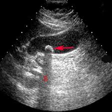
*Shadowing is a high attenuating artifact. Shadowing from a gallstone.
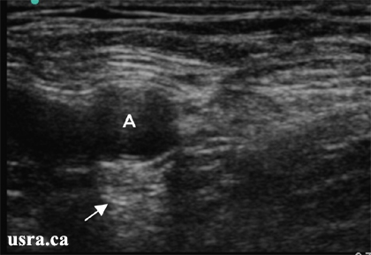
*Low attenuation structure results in an enhancing artifact (more echogenic distal to the structure). There is also a refraction artifact in this image causing an edge shadow.
Set your image depth so that the structure you want to image takes up 7/8 of the screen. If the image of the organ of interest takes up less screen space than that you are wasting the real estate of your screen.
Set your gain so that the image echogenicity is consistent from top to bottom of the screen.
Always scan a structure in two planes. Identify the boundaries of the structure you are interested in.
Lambert 7UP Scan
Mike credited Chris Kerwin with inventing the term 7UP scan.
The 7UP scan is a rapid ultrasound evaluation for the patient in shock.
It is a similar protocol to the RUSH (Rapid Ultrasound in Shock) Protocol.
Editors note: It was easier for me to grab some images of the RUSH protcol off the internet then get 7UP images. My apologies to Mike and Chris.
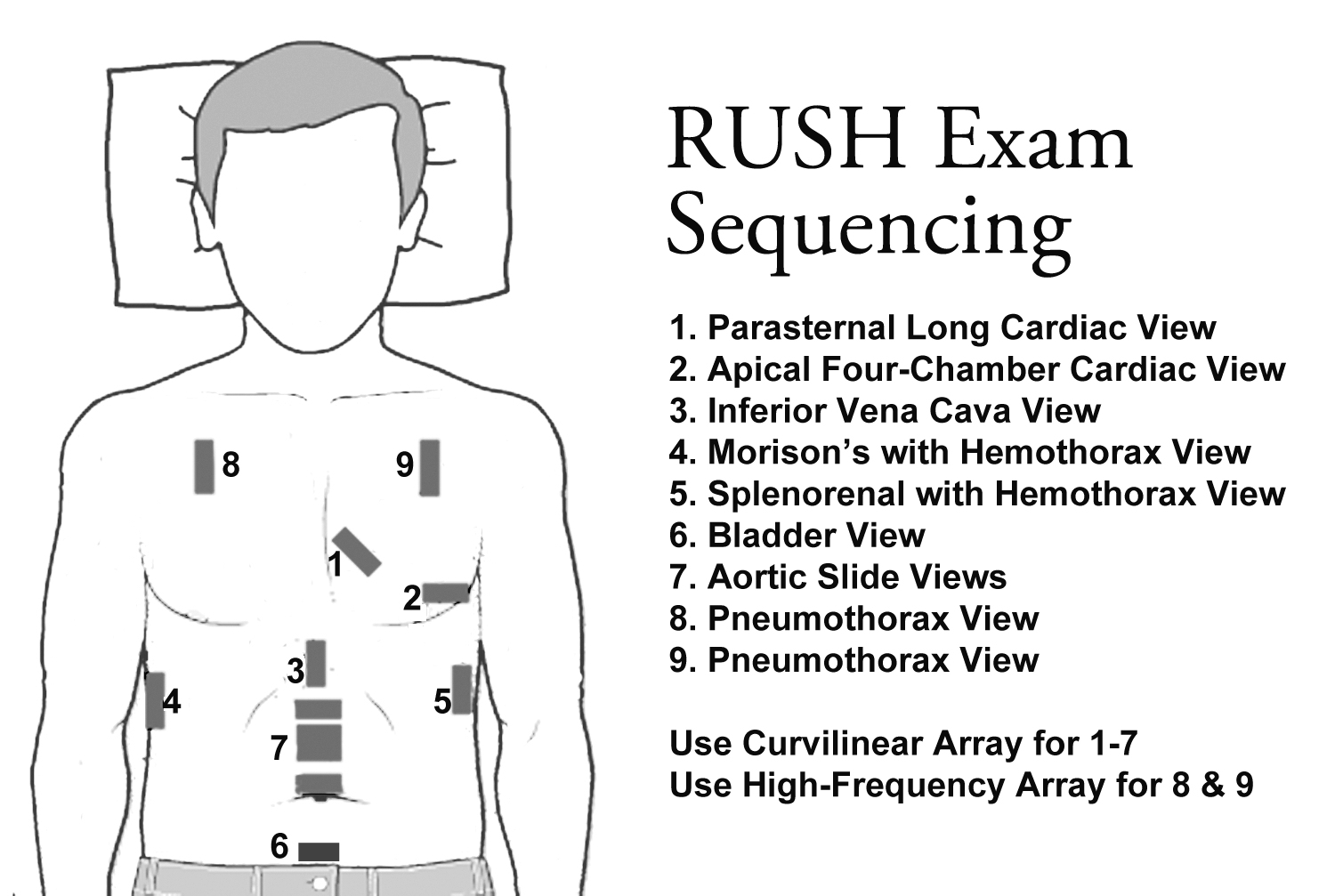
*RUSH Protocol

*RUSH Algorithm
The 7UP protocol also images the RUQ, Subcostal area, Abdomen, Pelvis, then Chest.
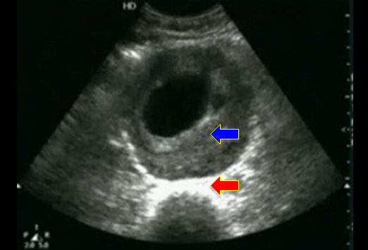
In a hypotensive patient with abdominal pain, fluid in Morrison’s pouch and a positive UCG there is nearly a 100% likelihood of ruptured ectopic pregnancy.
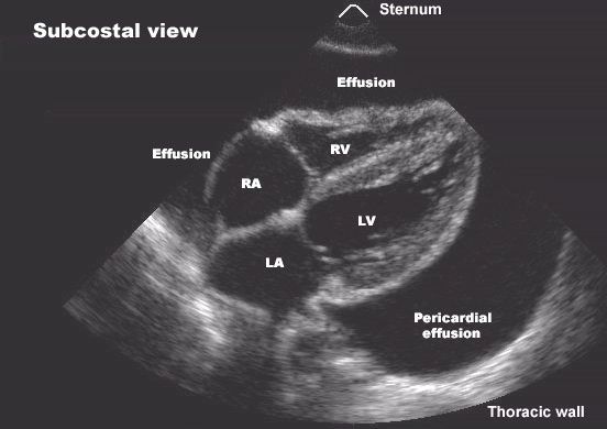
*Cardiac Tamponade with RV compression. Note the scalloped/compressed appearance of the RV.
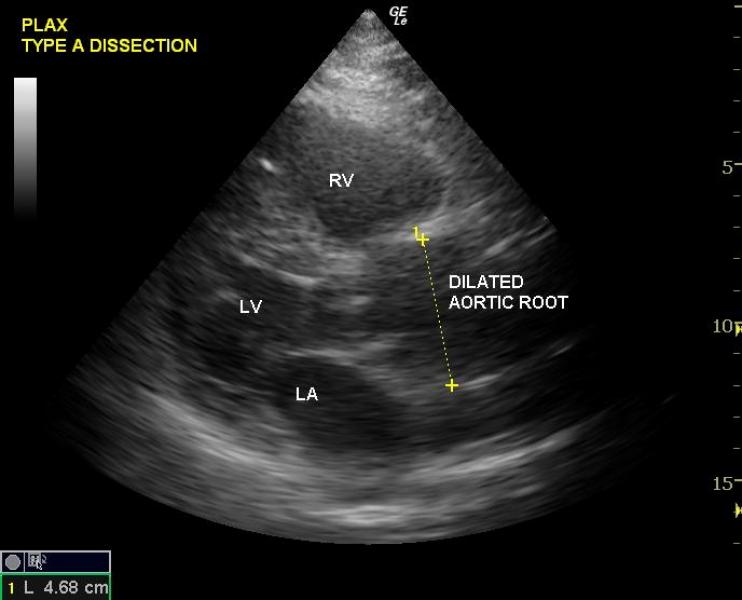
*Type A Aortic Dissection
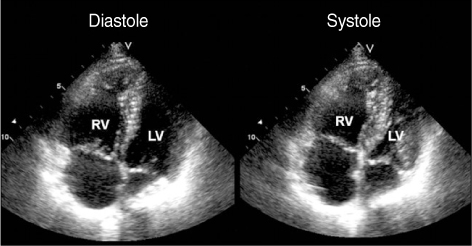
*Ultrasound consistent with PE. Typical RV is triangular in shape and smaller than LV. PE with increased right side pressures will show a proportionately larger RV.
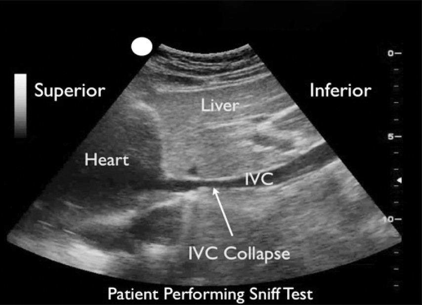
*IVC in Hypovolemia
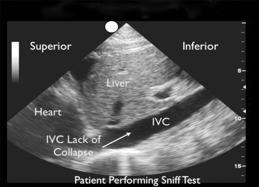
*IVC in Hypervolemia
*AAA with Thrombus
When looking for pneumothorax, image 3 sites on each hemithorax. If you don’t see normal sliding of the pleura, a pneumothorax is present.
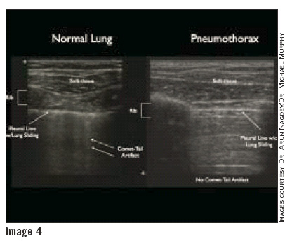
*Ultrasound Imaging of Pneumothorax. The left image is normal lung that shows pleural sliding and hyper-echoic comet tails. The right side image is a pneumothorax where pleural sliding and comet tails are absent.
Lambert/Frazer/Chastain/Chan and other Team Ultrasound Members Ultrasound Lab