
There are 6 images in this document. If you don't see them, scroll to the bottom of the page and click "view in browser".
Tekwani Study Guide
Inflamatory Bowel disease: Peak incidence is between ages 20-30. Patients will have abdominal tenderness. May have blood in stool.
Best management of uncomplicated diverticulitis: no ct scan and treat with oral antibiotics. Complicated diverticulitis: has phlegmon, perforation, stricture, abscess,etc, Ct is recommended if diverticulitis is suspected to be complicated by any of these
Ascending cholangitis due to obstructing stone: Treat with ERCP, IV fluids, IV broad spectrum antibiotics
If you find an anal fissure out of the midline, pt needs further consideration of HIV, cancer, crohn’s, syphilis, or foreign body insertion. Midline anal fissure is the most common cause of painful rectal bleeding. 90% are posterior and 10% are anterior. Midline anal fissures are due most commonly to hard stool.
Amebic liver abscess: entamoeba histolytica. Tx with hi dose metronidazole, think of it in patients from AZ or mexico or other endemic areas. Develops from eating food contamintated with feces. Pt’s present clinically about 8 weeks or more after travel. They have RUQ pain and fever.

Amdebic liver abscess
If patient has conjugated hyperbilirubinemia think obstruction of biliary tree: stones/atresia/sclerosing cholangitis. If ptatient has unconjugated hyperbilirubinemia, think of things that cause red cell destruction: hemolysis/sickle cell disease
Intersphinteric abscess develops from an anal gland obstruction and is the most common cause of anal fistula.

RLQ tenderness has the highest Liklihood Ratio of clinical findings for the diagnosis of appendicitis.
Work up for appendicitis in pregnancy: CT of abdomen/pelvis in the first trimester doubles the risk of childhood cancer in the fetus. MRI is an option but don’t give gadolinium in the first trimester. Most common option is to consult surgery for serial exams.
Acute ETOH hepatitis: enlarged liver and AST,ALT both elevated with a AST/ALT ratio greater than 2.
Worldwide, the most common cause of acute liver failure is APAP overdose. Most common cause of chronic liver failure is hepatitis.
Volvulus: 75% are sigmoid. Sigmoid are seen in elederly/bedridden patients. Can treat sigmoid volvulus with colonoscopy. 25% of volvuli are cecal. Seen in marathon runners/young patients. Treatment is surgical.
Initial treatment for toxic megacolon is fluids, antibiotics and steroids.
Hepatitis A never causes chronic hepatitis
IBS is more common in women, pain relieved by defecation. It is not a psychiatric disease.
Most common cause of large bowel obstruction is neoplasm.
Most common cause of massive lower GI bleeding is diverticulosis. Diverticulosis also most common cause of lower GI bleeding in general.
Lipase is more specific than amylase for pancreatitis. Both have similar sensitivities.
Toxic megacolon more common in UC. Perianal complications more common in Crohn’s disease
Schroeder Visual Diagnosis
Clinical diagnosis of anemia in dark skinned individuals: check the conjunctiva and palms. Palms will be more pale and less pink in the anemic, dark skinned individual.
Febbo question: Do we need to get written consent from patients to send a picture of an injury/illness to a consultant with our phones. Mistry/E. Kulstad response: No. Just get verbal consent from patient prior to sending photo to consultant. Be careful to limit any identifying info. Elise comment: Only do this for patient care. Do not send any images just because it is a cool or interesting picture. You can end up in a lot of trouble. Girzadas comment: Document pt’s verbal consent in the medical record.
Pitariasis rosea: Believed to be a viral illness. Rash lasts about 3 months. Kelly comment: Latest research is mixed on contagiousness. Rash is benign. Benadryl can be helpful for itching. Sun exposure helps relieve rash.
Gutate psoriasis: Small red patches with scaley surface. Gutate means” drop” and patches are raindrop size. This rash can follow strep infection by 2 weeks in genetically susceptible patients. Sun improves this rash.
Internal/external hordeolums can be treated with warm compresses and topical antibiotics.
Cradle cap: Treat with baby oil and combing out the flakes. If more severe, you can use lotrisone topically. Rash is greasy/oily appearing rash. As a contrast, eczema has a dryer appearance.
Numular eczema: coin shaped eczema in middle aged persons. This is more common in atopic persons or persons with hx of asthma.
Strep throat/scarlet fever: strawberry tongue with papillae and palatal petechiae. Scarlet fever has sandpaper appearance. Discussion about bicillinLA: Consensus that treatment with bicilin LA was totally acceptable if parents/patient prefer or if you are concerned about patient compliance.
Infected ear pits usually need surgical drainage. Can start with oral antibiotics and arrange surgery with ENT. Cover MRSA. Elise comment: don’t pop or drain in ED. It will be ineffective. There is some possible association between ear pits and renal malformation. There was consensus in the room that if you diagnose this, you don’t need to get a renal ultrasound on the patient.

Potts puffy tumor has mid forehead swelling. Admit for IV antibiotics. There is risk of intracranial extension. Infection most commonly involves anterior wall of frontal sinus. If toxic appearing get CT head to look for intracranial extension. If not toxic appearing, ok to admit with iv antibiotics and plan for mri of brain.

Potts puffy tumor
SSSS usually causes kids to be more uncomfortable/febrile/toxic vs impetigo where the kid is usually nontoxic/happy
Kerion: boggy inflammatory mass on scalp due to tinea capitus. Requires oral antifungals. Bill just starts griseofulvin and does not draw baseline labs. Diflucan is second line treatment. Don’t excise or drain.
Kawasaki’s conjunctivitis has perilimbic sparing with no drainage. Classic mimics of kawasaki’s are scarlet fever and adenovirus.
Sublingual swelling can be from a mucocoele or rannula. Requires elective excision.
Alopecia areata is well circumscribed hair loss.
Erythema multiforme: classically due to herpes, mycoplasma, medications. Rash may have target appearance. Elise comment: We see this very commonly. It is rare to progress to Steven Johnsons. Don’t over-react to this. Bill comment: if pt is on a sulfa drug stop it. Consider admission or good instructions to parents to watch for oral lesions or skin sloughing. Consider acyclovir for patients with cold sore or exposure to child with cold sore because herpes infection is more likely with this clinical scenario. Bill does not use steroids for this rash.
Ryan Med Student Elective Update
Heller Pediatric Abdominal Radiology
Consult your imaging consultants if you have uncertainty about the best imaging study to answer a clinical question.
Neonatal bowel obstruction: Clinical exam cannot differentiate between high and low types of obstruction. Bilious emesis is a marker of malrotation. However, bilious emesis in a child less than 36 hours old doesn’t always mean malrotation. Begin imaging with a plain abdominal xray in the child under 36 hours long. Double bubble sign is almost always duodenal atresia and requires surgery.
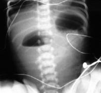
Double bubble sign
Upper obstruction can be due to Malrotation that lead to midgut volvulus. Bilious emesis in the newborn who has been normal for a few days should be considered midgut volvulus until proven otherwise. Elise comment: no need to do a plain xray in the previously normal newborn with bilious emesis. This child has likely midgut volvulus and should go right to upper GI. Non-bilious emesis in 4-8 week child, think pyloric stneosis. In a normal child you can’t even find the pyloris on u/s. Harwood comment: what do you do with kids with borderline U/S findings for pyloric stenosis? Heller reply: We re-image in 48 hours. If child dehydrated or not feeding, then admit. If hydrating OK then child can go home and get second U/S as outpatient.
Low neonatal obstruction usually shows numerous gas filled bowel loops on KUB. Low obstructive causes: imperforate anus, hirschprungs, small left colon, meconium ileus, ileal atresia. Work up for low obstructions is barium enema. Meconium ileus is thick meconium obstructing the bowel. Associated with CF. Hirschprungs is due to an absence of ganglion cells in rectum. Pt’s have a low rectal/sgmoid ratio. Basically the rectum is narrower than sigmoid. Associated with downs syndrome.

Low Obstruction from Hirschprung's Disease
Adhesions/appy/inguinal hernia/intussusceptions/malrotation/meckel’s AAIIMM mnemonic for SBO.
Intussusception: 90% are ileo-colic. 90% are idiopathic. Ideopathic means lymphoid hyperplasia is thought to be causing intussusception. Sweet spot for age for intussusceptions is 6mo to 2 years. More common in winter/spring. Initially patients will have crampy abdominal pain. If this progresses without identification over a couple of days then kids become lethargic. Start with KUB. Second step is U/S. U/S should be able to diagnose or exclude the disease. Heller felt that U/S is a good test for intussusception. Treat with enema reduction (air or liquid contrast). Younger radiologists prefer air contrast enemas. Barium for enema is risky because if there is a perforation it causes peritonitis. Gatrograffen is also risky because it is hyperosmoler. If there is a perforation it can cause life-threatening electrolyte abnormalities. Radiologists dilute gastrograffen 5:1 with water to reduce osmolality. Air contrast enema does not carry an increased risk of perforation. You have to have surgery on alert prior to doing a barium enema.
Appendicitis: Start with U/S. 2nd test is an appendicitis protocol CT. This is a pelvic CT with IV and enteric (oral or rectal) contrast. Heller’s comment is if you are going to expose the child to radiation, take your best shot with both IV and enteric contrast. Herrmann comment: sometimes this study can miss the appendix. Heller reply: You see the appendix 99% of the time with this study. Non-visualization of the appendix and secondary signs of inflammation is reliable evidence that makes appendicitis much less likely. Most common missed diagnosis with limited appendicitis protocol is pneumonia.
Radiation safety: Lifetime Risk of fatal malignancy from CT is approximately 1:1000.