Conference Notes 9-3-2013

Reminder  Emergency Medicine Foundation Golf Outing Tuesday afternoon/evening, October 8th.      

Gupta     Study Guide     Trauma

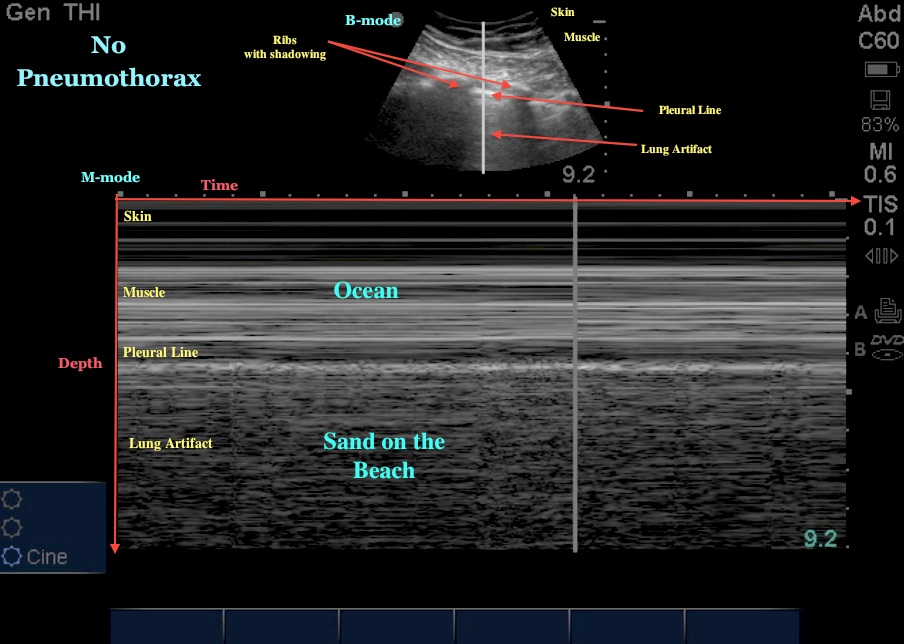
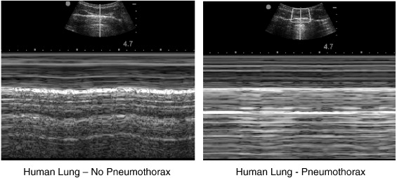
If you suspect tension pneumothorax, do immediate needle thoracostomy.  Do not wait for CXR or a clinical response to IV fluids.

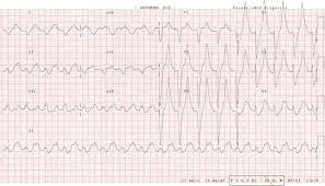
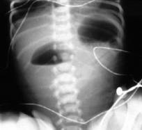
If you are seeing this CXR you are late in needling the chest:

CT is more sensitive/specific than ultrasound for diagnosing solid organ injury.   It is more sensitive than ultrasound for picking up small amounts of intraperitoneal blood. CT is not great for picking up hollow viscous injuries.    Harwood comment: Use serial exams to diagnose hollow viscous injuries if you are suspicious and CT is non-diagnostic.

Kelly Williamson comment:  In adults with blunt flank trauma who don’t have gross hematuria and who are not hypotensive, you don’t need to work up the patient for renal injury.   You don’t need to send the urine to the lab.   You can visually inspect the urine to check for gross hematuria.  If the urine is not grossly bloody, you don’t need to work patient up for renal injury.

In contrast to adults, microscopic hematuria in pediatric patients who suffered blunt trauma still requires further evaluation.  Harwood comment:  You won’t find a lot of treatable injuries using microscopic hematuria as your only criteria to do a IV contrast CT.    Gupta comment: For the boards,  microscopic hematuria in adults requires no further urinary tract evaluation.   Microscopic hematuria in kids,  requires a CT abdomen/pelvis with iv contrast.  Elise comment: The injury you would be looking for is at the junction between the kidney and the ureter.  This area is very mobile in kids and is prone to injury.    Harwood/Elise/Erik/Christine comment: They wouldn’t CT a kid for just a few RBC’s in their urine.   The kid would need in addition some history or sign of serious trauma to proceed with CT.  There was then a discussion about the recent publication of the PECARN decision instrument to decide on work up of the abdomen in kids with blunt trauma.


Elise comments: Strengths-as always with PECARN, really big study, well done, reliable results. Weakness- they don't incorporate US in their workup (during EMRAP interview with an author he stated that some centers weren't using US routinely).  Doesn't specifically discuss microscopic hematuria, but simple 7 point instrument to help decrease CT use in these kids.  Decision instrument missed 2 kidney injuries:  one kid had gross hematuria, the other had distracting injury (rib fracture) and microscopic hematuria (10 RBC per hpf).

Harwood comments: This is a great rule. It has a 99.9% (-)  predictive value & doesn't use micro urine or LFT's (in old rules). It did missed 7/203 injuries (requiring treatment). However, of the 7 misses, 2 kids were 2 y/o's. 1 did indeed have gross hematuria & was treated without an operation (did get a blood transfusion). 5 were 16-17 y/o's. All would have gotten ED CT's based on presentation. This includes a 17 y/o MVA had rib injury, blood in the belly (& 10 RBC's in urine). This pt got got embolization (spleen? most likely).


Intraperitoneal bladder rupture usually results from a compressive force to abdomen causing rupture of dome of the bladder.   Gross hematuria is present 98% of the time.  


Extraperitoneal bladder rupture is caused usually by sharp boney edges of pelvic fracture.


Large myocardial contusions can result in cardiogenic shock.   Harwood and Girzadas comment: There is no good diagnostic test for myocardial contusion and there is no accepted treatment for myocardial contusion so there is no point expending resources looking for this.   If the patient is in shock you gotta think hemorrhage, hemorrhage, hemorrhage.  But if hemorrhage is ruled out and the pt is hypotensive or has an arrhythmia it could possibly be from myocardial contusion but still is more likely to be due to hemorrhage.

The spleen is the most common  organ injured in blunt trauma . Blunt trauma; organs most frequently injured in decreasing order: Spleen>liver>kidney>small bowel .  Penetrating trauma; organs most frequently injured in decreasing order:  Small intestine>liver>colon.      

Harwood comment:  In elderly patients with multiple rib fractures (3-4 broken ribs) , they have a surprisingly high mortality.   Admit them for observation.  Christine comment: Mortality in the elderly increases by 19% for every rib broken above 2.

Left sided diaphragmatic hernias are three times more likely than right side.   The liver is thought to protect the right diaphragm.  Diaphragm injuries can be subtle initially and pt may become symptomatic years later.

Treat patients with pelvic fractures and hypotension with  pelvic binding, prbc/ffp transfusion,  and angiography with embolization.

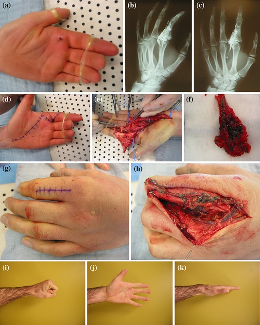
Jakubovich     Pediatric Trauma   M&M

Pediatric GSW victim(5yo) transferred from outside hospital to ACMC.  Intubation was attempted at initial hospital but was either unsuccessful or tube became dislodged.    Upon arrival at ACMC,  pt could not be intubated using laryngoscopy.   Resident and attending were also unable to intubate with glidescope.   Patient, however, was able to be bag-valve-mask ventilated.    ED/Surg team proceeded to perform cricothyrotomy on pt.  PRBC transfusion started.

Pt went to OR and had multiple intra-abdominal injuries repaired.   In the OR, the cricothyrotomy developed an air leak and was removed.  Peds annesthesia was able to intubate the patient.

Patient was discharged home.  He suffered from PTSD.

Airway issues:   In infants with prominent occiput, you can put a towel under the shoulders to line up the airway.    In kids age 5 or older, it is recommended to put a towel under the head to line up the airway.  Harwood comment:  Place a towel under the shoulders first, if that doesn’t work to line up the airway, move the towel to behind the head and look again. 

Pediatric bougie will fit through a 4 ET tube.   Adult bougie will fit through a 5.5 ET tube.

Since we were able to bag-valve-mask this patient, did we need to cric this patient?  Elise comment: A pediatric LMA may have been a reasonable option in this situation.  Our pediatric difficult airway box should have a pediatric sized LMA. Erik comment:  Maybe a retrograde intubation over a wire passed through the cric would be a nice technique.  Kelly comment: Don’t beat yourself up over doing the cric, you need an airway and getting the airway anyway you can is OK.   Christine/Elise comment: If you can bag the patient successfully, you usually have time to consult pediatric anesthesia emergently.   There was some consensus among attendings present that an LMA would have been the optimal bridge device until pediatric anesthesia could be consulted.

Should we have done an open cric or a needle cric?  Age recommendations go down to 5yo for open cric.  However at age 5 and below it will be more difficult due to anatomic size.   In this case Trauma Surgery had no difficulty passing a shiley through the cricothyroid membrane.   Harwood: It is best to use a commercial Melker percutaneous seldinger technique cricothyrotomy kit.   Other jerry-rigged needle cricothyrotomy techniques and jerry-rigged jet insuflation are prone to failure.  Elise: I agree the Melker kit is the best option.  But again, if you can ventilate a patient it is better to continue bagging or place an LMA and get help from anesthesia.   Christine comment: Your respiratory therapists will have no experience with needle crics.   Erik comment: It may be helpful to  remember that in many places around the world, they do entire surgical procedures without intubation and just bag-valve-mask the patient.

Elise follow up comment: We currently stock size 3 LMA in the PED.  This is sized for patients weighing 30 - 50 kg, and would work for average sized kids age 8 to small adult.

Plan is to add smaller sizes (2 and 2.5) which will cover kids 1 year old and up.  There are smaller LMAs, but my thought is to keep this simple, and little babies are usually less challenging intubations.  

Getting blood:  You can make an MR# for a DOE name prior to arrival of the patient to order blood from Blood Bank.

Kmetuk        Cardiac Trauma

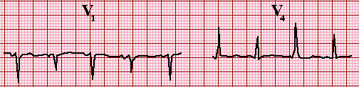
Case 1.    Penetrating left chest trauma.   CXR discussed by Harwood shows hemothorax despite left chest tube, heart is enlarged and is very suspicious in a young patient for hemopericardium.   FAST exam identified hemopericardium.   Pt went to OR for repair of RV.  Pt did well.

Cardiac Box  

RV is the most commonly injured cardiac structure due to penetrating trauma.  Penetrating injury to the chest can definitely injure abdominal organs as well.   Left ventricle is more likely to self seal than right ventricle.   Stab to the heart is 17X more likely to survive than GSW to the heart.

Salzman comment: Patients with stab wounds to the heart  are the one’s who can survive an ED thoracotomy.    If you do an ED thoracotomy, push the ET tube down the right mainstem bronchus to deflate the left lung and improve your visualization of the heart.

Salzman comment:  If you have pressors infusing in a young trauma patient it means they need blood Dammit!   Young trauma patients should never get pressors (except possibly neurogenic shock) they need blood.

Salzman comment: You can close wound in the RV with foley catheter, stitches, or staples, or just having someone put a finger in the hole.

Salzman comment:  In a 20something patient with penetrating chest trauma, and FAST shows some pericardial fluid, that is blood.    Also in a patient who is arrested or peri-arrested don’t hesitate, just get started on the thoracotomy.  ( As an aside, when breaking bad news to families, in a similar fashion get to the fact that their loved one is dead with little preamble. )  There is no role for needle pericardiocentesis in penetrating cardiac injury.   The blood in the pericardial sac is usually clotted and can’t be aspirated with a needle.

Case 2.  51yo male victim of motorcycle vs. truck collision. Blunt chest trauma.  Intubated, hypotensive.   CXR shows wide mediastinum.    CT chest shows aortic injury.   PT was started on esmolol but BP dropped and esmolol was stopped.  Pt’s aorta was repaired in the OR with a graft.   Pt walked out of hospital 20 days later.

90% of patients with aortic injury will die at the scene.  50% of the survivors die in the next 24 hours.

High risk historical factors for aortic injury: rapid deceleration injury, t strikemvc, fall more than 3 stories.

Febbo question:  Do you need to CT scan an asymptomatic patient who has one of the above mechanisms?    Salzman comment: Yes, he has seen pt’s who have intimal injuries or pseudoanuerysms that have been relatively assymptomatic. 

Walchuk       Mandibular Dislocation

Anterior dislocation is the more common dislocation and is reducible in the ED.   Posterior dislocations are rare and need specialty consultation and likely OR reduction/surgery.

Pt’s with anterior dislocations can’t close their mouths.  There will be a preauricular depression in the skin.   Check cranial nerves 5 and 7.   Unilateral dislocations will have jaw deviation to the side opposite the dislocation.

Posterior dislocations can result in external auditory canal injury.

Intra TMJ lidocaine can give significant pain relief and even allow reduction.

You can try to elicit a gag response.  The gag response blocks contraction of the masseter and can result in reduction of the dislocation.

Dan Gromis method of masseter massage was presented on video.    Basically, an assistant massages the masseters on both sides while the operator applies inferior and posterior pressure on the mandible while at the same time tilting the chin upward.   The video demonstrated very easy and non-painful reductions.

After reduction, pt should be on a soft diet for a couple of days.

Harwood comment: Main thing is to apply some downward traction on the mandible and get the chin elevated relative to the angle of the mandible and then move the mandible posteriorly.

Lam       Fever in infants 0-90 days

Fever/age cutoffs to initiate work up in kids:  0-90 days=38C    3mo-36mo=39C

10% of kids 0-90 days with 38C or higher temp will have serious bacterial illness.

Clear cut national consensus to do sepsis workup and admit/give IV antibiotics in kids 0-28 days.   Give Amp and Cefotaxime.   Sam said he has the experience of regularly giving  ceftriaxone in kids 0-28 days with no complications during his fellowship.

Well appearing infants 29-90 days old with Temp 38 or higher is the difficult group to make decisions on.

 Rochester criteria: WBC 5-15K, UA with less than 10wbc/hpf, less than 5wbc in the stool.  No LP criteria.  Also kids can’t have any significant pmh or clinical exam findings of serious infection.  These low risk criteria miss 15-45% of serious bacterial illness.. 

Boston criteria: WBC under 20k, CSF with less than 10wbc, UA with less than 10 WBC or neg urine dip. All kids got 50mg/kg ceftriaxone IM.  All had re-check and second dose of ceftriaxone in 24 hours.   These criteria missed 6% of serious bacterial illnesses.

Philadelphia criteria: Non-toxic, WBC <15K, low bands, urine with <10wbc /hpf and no bacteria on gram stain, CSF<8wbc/hpf, CXR with no infiltrate,  Stool with not a lot of WBC’s.  High risk kids were admitted with antibiotics.  Low risk kids did not receive antibiotics and were sent home.  This strategy missed the least amount of serious bacterial illnesses.

In this age group there is a 10% chance of viral meningitis.

If you are going to treat a febrile child in this age group with ceftriaxone, you need to do a LP prior to giving ceftriaxone.   There is national consensus on this management point as well.

OM does not make a child in this age group low risk.  They still need an LP.

Bloody taps require admission and antibiotics because the results are uninterpretable. 

Sam uses the Philadelphia criteria in kids less than 8 weeks.  He does a full septic workup on them.  If results fit low risk criteria, he sends them home.   Scott Altman and Sam both said they will give kids antibiotics (ceftriaxone) prior to sending them home.  

There was some practice variability voiced in the group.   Some faculty will do full septic workup, give antibiotics, and admit for observation in all these febrile kids 0-8 weeks. 

Occult bacteremia in kids 3 months to 36 months in the era of pneumo and HIB vaccinations is basically dead.  The incidence of contaminated blood cultures is approximately 10X the incidence of positive blood culture. So getting blood cultures in these patients leads to many more false positives than true positives.   In Immunized kids 3-36 months with high fever, instead of getting blood cultures, look for uti.  A kid with 2-3 doses of vaccine can probably be considered immunized.

In kids in this  age group, if you get a CBC for whatever reason and the WBC is over 20K get UA/UCX and get a CXR.  Kids with a WBC >20 are at higher risk for both.

Kids who have a clear cut diagnosis of a viral illness(positive rsv, chicken pox, croup, bronchiolitis,etc), meningitis is very unlikely.   

Thanks for reading!  Stay safe out there.   Double glove and be cautious/take your time with procedures to avoid Sharps Injuries.  


Conference Notes 8-20-2013

Some Awesome Pearls at Conference this week!  But first gotta promote the Foundation:

SAVE THE DATE for the ACMC EM Foundation Golf Outing

What: Emergency Medicine Golf Outing – October 1, 2013

 Location: TBD

Time: Following the Emergency Medicine Conference/Early Dept Meeting and Vice-Chair Vote

Cost: $150/golfer (includes golf, dinner & drinks)

Purpose: To raise awareness for the Emergency Medicine Endowment which provides funding for residents to further their education

Contact: Jason Keene, Director of Development 708-684-2012 or jason.keene@advocatehealth.com

*Proceeds will benefit the Emergency Medicine Endowment

*Electronic Invitation to follow


McKean/C. Kulstad     Oral Boards

Case1.   TTP    Critical Actions: plasmapheresis (the same thing as plasma exchange) is the key treatment, CT head, No LP, Admit to ICU, get Hematology Consult.  Optimal care: DIC panel, discuss FFP transfusion, assess for life threatening bleed,  discuss steroids with hematology.   Classic TTP Pentad:  microangiopathic hemolytic anemia (MAHA), neurologic abnormalities, fever, renal disease, thrombocytopenia.  Frequently the pentad is not present.  The cornerstones of the diagnosis of TTP are thrombocytopenia and hemolytic anemia.  Most patients have some neurologic and renal abnormalities but the severity of the neuro and renal abnormalities is highly variable.     This disease is more common in middle age persons. Females more commonly affected than males.  TTP has high mortality  90% untreated and 20% treated.   Don’t give platelets.  TTP and HUS are very similar disease processes in adults.

Group discussion about management of TTP in regard to LP.   Most agreed when there is diagnostic uncertainty between TTP and meningitis give antibiotics and steroids prior to getting labs back and prior to CT/ LP.  If platelet count is less than 50K (Harwood’s cutoff) don’t do LP.  If platelets above 50K probably safe to do LP.  Girzadas: Some case series have shown no bleeding complications with very low platelet counts.    Erik comment: If you do a LP for possible meningitis and the platelet count comes back below 50K, the patient will likely be fine.   Everyone agreed though that Harwood’s cut off at 50K was prudent.

Up to Date reference:  It is traditionally stated that a platelet count greater than 40,000 to 50,000/microL provides safety for interventional procedures such as lumbar puncture [11-13]. In certain procedures where bleeding risks are greater, or the risk of complications from minor bleeding is high, a platelet count greater than 80,000 to 100,000/microL is often required by surgeons (especially neurosurgeons) or anesthesiologists.

A quick Abstract on this topic:

The risk of spinal haematoma following neuraxial anaesthesia or lumbar puncture in thrombocytopenic individuals.

van Veen JJ, Nokes TJ, Makris M.

Source

Sheffield Haemophilia and Thrombosis Centre, Royal Hallamshire Hospital, Sheffield, UK. joost.vanveen@ukgateway.net

Abstract

Neuraxial anaesthesia is increasingly performed in thrombocytopenic patients at the time of delivery of pregnancy. There is a lack of data regarding the optimum platelet count at which spinal procedures can be safely performed. Reports are often confounded by the presence of other risk factors for spinal haematomata, such as anticoagulants, antiplatelet agents and other acquired or congenital coagulopathies/platelet function defects or rapidly falling platelet counts. In the absence of these additional risk factors, a platelet count of 80 x 10(9)/l is a 'safe' count for placing an epidural or spinal anaesthetic and 40 x 10(9)/l is a 'safe' count for lumbar puncture. It is likely that lower platelet counts may also be safe but there is insufficient published evidence to make recommendations for lower levels at this stage. For patients with platelet counts of 50-80 x 10(9)/l requiring epidural or spinal anaesthesia and patients with a platelet count 20-40 x 10(9)/l requiring a lumbar puncture, an individual decision based on assessment of risks and benefits should be made.

Case2.   Achilles tendon rupture    Critical actions: Complete extremity exam,  consult ortho, splint in moderate  plantar flexion.  Tear is usually just above the calcaneus.   Surgical repair has less risk of re-rupture but of course carries surgical complications.   Plantaris tendon rupture causes whip like pain in calf.  Treatment of plantaris rupture is pain control  and ace wrap.  Medial gastrocnemius tear also can cause sudden calf pain and is treated with conservative measures.

Thompson's Test: Right leg in photo is abnormal and doesn't plantar flex with squeezing the calf (positive test=tendon rupture). You can also do the test with the patient prone and knee flexed.

Discussion between Harwood and Elise about best position to splint ankle with Achilles tendon rupture.  Harwood basically said it is not that important to splint in plantar flexion.  Elise felt plantar flexion de-stressed the tendon and was  preferred over  splinting  at 90 degrees.

Case3.   Perforated Ulcer    Critical actions: IV antibiotics (cover gram negatives & anaerobes)  and emergent surgical consult.    Optimal care: PPI, NG, foley. 

Free air under the diaphragm on CXR:


C. Kulstad    Study Guide Trauma

Nexus

The NLC decision instrument stipulates that radiography is not necessary if patients satisfy ALL five of the following low-risk criteria:

  • §  Absence of posterior midline cervical tenderness
  • §  Normal level of alertness
  • §  No evidence of intoxication
  • §  No abnormal neurologic findings
  • §  No painful distracting injuries


Canadian C-spine rules

The CCR involves the following steps:

  • §  Condition One: Perform radiography in patients with any of the following:
    • ·         Age 65 years or older
    • ·         Dangerous mechanism of injury: fall from 1 m (3 ft) or five stairs; axial load to the head, such as diving accident; motor vehicle crash at high speed (>100 km/hour [>62 mph]); motorized recreational vehicle accident; ejection from a vehicle; bicycle collision with an immovable object, such as tree or parked car
    • ·         Paresthesias in the extremities
  • § 
    • ·         Simple rear end motor vehicle accident; excludes: pushed into oncoming traffic; hit by bus or large truck; rollover; hit by high speed (>100 km/hour [>62 mph]) vehicle
    • ·         Sitting position in emergency department
    • ·         Ambulatory at any time
    • ·         Delayed onset of neck pain
    • ·         Absence of midline cervical spine tenderness

Patients without any of the low-risk factors listed here are NOT suitable for range of motion testing; they must be assessed with radiographs.

If a patient does exhibit any of the low-risk factors, perform range of motion testing, as described in Condition Three below.

  • §  not

Head injury on Coumadin: Check INR,  observe in ED for a few hours   Observe  in hospital if INR is very high. 

 Pregnant patient with chest trauma and pneumothorax: Place tube in the 3rd intercostal space in the mid-axillary line.   It’s alittle higher placement than in a non-pregnant patient.

In contrast to orbital floor “blow out fx’s”,  Orbital roof fractures and fractures through the naso-ethmoid bone risk injury or infection to the brain.  These roof and naso-ethomid fractures need admission and consult of neurosurgery.

Stable C-spine fractures: compression less than 25%, spinous process fracture, transverse process fracture.

 Fractures of spinous and transverse processes are stable.


Treatment of severe blunt laryngeal injury (laryngeal fx).   Group discussion on this one:  Start with RSI but be prepared to have great difficulty with the airway.  You want to paralyze the patient to give you an optimal look but loss of muscle tone may cause the airway to collapse.  There is a high risk of creating a false passage with the ET tube.  Your rescue approach is problematic as well.  Cricothyrotomy is not recommended due to disruption of the normal cricothyroid anatomy.   Harwood and Kelly Williamson suggested being prepared to do a seldinger cric through the trachea in this situation.   Everyone cringed at facing this difficult situation.

Zones and Triangles of the neck


Girzadas comment: Way to remember that zone 1 is at the bottom and zone 3 is at the top. Big Red 1= Aorta is in zone 1 and Zone 3 is up toward the Third ventricle. 

Open mandible fractures require admission for IV antibiotics.   Open fracture is identified by blood between teeth.   Tongue blade test is 96% sensitive for identifying mandible fracture.   Closed  mandible fractures can go home.

NG and foley sizes for kids are 2X the ET tube size.   Chest tubes are 4X the ET tube size.  ET tube size is age/4 + 4.

Evaluate for incomplete cord injuries with anal wink, cremasteric reflex, and bubocavernosis reflex.

Chest tube output to prompt OR in Kids: 15ml/kg initially and 4ml/kg/hr ongoing.  For adults: 1500ml initial and 200ml/hr x 4 hours

Central cord syndrome: Upper extremities weaker than lower extremities and proximal weakness more pronounced than distal weakness.  Man in a barrel is the classic analogy for this neuro deficit. 

Brown Sequard syndrome : Ipsilateral paralysis and loss of pain and temperature on the contralateral side.


Subdural/Epidural/SAH Bleeds


Williamson/Patel/Tekwani         Post-Partum Emergencies

Case 1: 36 yo female G2P2 s/p c-section one week ago.  Pt had headache.  She then developed some chest pain.   BP 132/80  P 52  P/O 98%.    Broad ddx of head and chest emergencies was considered.    Kari was thinking pt had predominantly a headache problem.  Pt improved with treatment.   D-dimer was 17.  CXR was pretty much pristine.   CT-PE study showed aortic dissection extending to innominate and carotid arteries.    There was debate between radiology and CT surgery about whether the CT findings were artifact.    Kari called in Cardiology to do transesophageal echo.  TEE showed/confirmed  dissection.  Pt went to OR.   Discussion about CT evaluation of PE vs. Dissection.   If you are specifically looking for one disorder (Dissection or PE) get the appropriate test.  If both are in the differential, get the CT PE. You are probably more likely to see a dissection on the PE study than see a PE on the dissection study.

Kari comment:  The presentation of dissection can be extremely protean or subtle.

Case 2: 38yo female with chest pain 9days after c-section.     DDX was PE, dissection, musculoskeletal pain,  pre-ecclampsia, nstemi, cardiomyopathy.  EKG showed subtle inferior depression and high lateral minimal ST elevation.  Most people in the room read it as nonspecific.  Nobody would have called a STEMI on this EKG.  Troponin was elevated.   Pt got cathed and was found to have LAD dissection.  There are multiple case reports of post-partum woman having spontaneous coronary artery dissections. 

Coronary artery dissection or thrombosis is more common in the post-partum patient than acute coronary syndrome.  These patients don’t usually have underlying atherosclerosis.

Case 3.   31 yo female 1 week after delivery.   Pt has sob.  Went to an urgent care center the day before.   Pt had nl ekg and neg d-dimer, trop from previous visit at urgent care center.   DDX=PE, post-partum cardiomyopathy, pericardial effusion, anemia, pre-ecclampsia.  EKG did not show any acute ischemia.  CXR is pristine.  Trops are negative.  Hgb is 11.6. VQ is low prob.  Venous dopplers are negative.   Ambulatory pulse ox was nl.  Pt had no protein in urine.  BP was 156/83.  Pt was dc’d home…..    Pt develops a headache 5 days later and she collapses.  Pt was severely  hypertensive.   She arrests in the ED. Difficult intubation.   CT head was neg.  Pt was placed on magnesium and labetalol.   She remained in a coma and support was withdrawn.  Diagnosis was pre-ecclampsia.

20% of patients present with htn alone (>140/90) and no proteinuria.  Ecclampsia can present up to 8 weeks after delivery.   It is critical to get a urine protein/creatinine ratio.  This can be ordered as a single urine test from lab.  Anything above 0.19 suggest pre-ecclampsia.  Also get CBC and CMP.  Treat pre-ecclampsia with magnesium.      

Lecturers comments:  The post-partum patient should be considered very carefully and deserves a more detailed work up.

#1 risk factor for endometritis is C-section.   Harwood comment: There is a sub population that can be treated as an outpt.    Kelly’s comment: If patients receive IV antibiotics in the hospital, when they clinically improve and defervesce they no longer need any antibiotics IV or oral.

Peripartum cardiomyopathy can go out to 5 months after delivery.  

PE is more likely post-partum than during pregnancy.

Harwood comment:  Pregnancy is a stress test.  Some women fail this stress test and get head bleeds, aneurysms, post partum cardiomyopathy, pe’s , and dissections.

Frazer       Safety Lecture

Sign out: Admit to MICU for ativan overdose.  Pt is somnolent but stable and is boarding in the ER waiting for the ICU to open up.   Pt has been in ED for several hours.   He becomes more alert.  He gets downgraded to step down unit.   Pt gets transferred up to floor.  Although the ICU attending was contacted, no one ever contacted the floor attending that the patient was going to the floor.  The patient spent several hours on the floor with no orders.

All attending should be contacted when there is an upgrade or downgrade of admission destination.

There was a trouble shooting discussion and the group felt that  putting the medical team as a consult with the reason as MICU admit.    At sign out we should be asking each other is this patient downgradable?   If so who needs to be contacted.

Felder       CV Workshop

It should be a “map” of your career and interests.  

Put  information lines under each heading  in reverse chronological order.

Keep it concise.    If you have a lot of pubs just list topics and the number of references under each topic.  Then add at the bottom: Detailed list of publications on request.

Make a top 5 list of achievements/strengths/interests.   Be sure your CV highlights these strengths.

Girzadas comment:  Use the decision points in residency to differentiate yourself on your CV.   Lectures, elective rotations, resident project, and leadership positions all can be used strategically to demonstrate a focus in your residency training. 

5 quick fixes:

  •  
    1. Creative editing like bolding should be used to highlight your strengths.
    2. Make  your career timeline easy to follow.
    3. List your publications but if you have many make it more conscise as described above.
    4. Make the CV easy on the eyes
    5. Make formatting consistent.

You may want to customize your CV to different jobs you are applying to.

Before you interview for your dream job, think about applying and interviewing for a job you are not super-interested in to give yourself a chance to practice.

Cover letters should give alittle more definition to who you are and what kind of doctor you are beyond the objective work milestones on your CV.    Christine comment:  It should give the employer some info about why you are interested in a particular geographic area or hospital.  

Altman comment:  Personal connections such as phone calls and networking are the most powerful way to get jobs.    Say,  “Dr. X told me to call you”


Ryan                       Medical Student Review

Conference Notes 8-13-2013

Another Outstanding Week in Conference!  but first an important word from our sponsor: 

SAVE THE DATE for the ACMC EM Foundation Golf Outing

What: Emergency Medicine Golf Outing – October 1, 2013

 Location: TBD

Time: Immediately Following the Emergency Medicine Conference

Cost: $150/golfer (includes golf, dinner & drinks)

Purpose: To raise awareness for the Emergency Medicine Endowment which provides funding for residents to further their education

Contact: Jason Keene, Director of Development 708-684-2012 or jason.keene@advocatehealth.com

*Proceeds will benefit the Emergency Medicine Endowment

*Electronic Invitation to follow


Lotsa pics in these notes if you don't see them scroll to the bottom and click "read in browser"

Coghlan      Study Guide   ENT

Most common source of bleeding in posterior nose bleeds is the sphenopalatine artery.

Compications of posterior nasal packs: eustacian tube dysfunction, nasopulmonary reflex  causing  apnea/bradycardia/hypoxia/bronchoconstriction/cardiac arrest,  necrosis of the columella. 

Best nerve block for complex ear laceration is the auriculotemporal block.


Diabetic with signs of otitis externa:  Consider malignant OE due to pseudomonas.   These patients are ill-appearing, diabetic, febrile, elderly and have granulation tissue in their external ear canal.


Malignant OE


OE with TM perforation:  Use Floxin because it is the only topical antibiotic approved by FDA for use with an open middle ear.    If cost is an issue, use Cortisporin otic suspension as an alternative .

Tooth avulsion: never re-implant primary teeth.  Adult tooth that is avulsed hold by crown,  irrigate with saline and re-implant in the socket ASAP.   Beyond 2 hours the chance of success of re-mplantation is quite low.   Best transport mediums for an avulsed tooth is Hank’s solution.   Saliva, milk, saline are 2nd, 3rd, 4th best..

ANUG (acute necrotizing gingivitis) :  Due to oral anaerobes,  pt’s have “punched out inter-dental lesions of the gingival”.    Treat with antibiotics that cover anaerobes.

    ANUG


Lemierres syndrome:  Infectious thrombophelbitis of the IJ due to a parapharyngeal space infection.  Pt’s can get septic pulmonary emboli.


Diagram of Lemierre's Syndrome


Aphthous stomatitis: Cellular immune mediated response.   Treat with pain medication and steroids.

Pt  gets hit in the mouth with a baseball.  Pt has dental injury.  Dental mobility but not displacement is called subluxation.    Dental mobility and displacement=luxation.    There can be intrusive, extrusive, posterior and anterior luxations

For burns, don’t use silvadene above the clavicles as it can permanaently alter the color of facial skin.

There was a discussion about complications from OM.   Harwood comment: This is a bogus question.  It is extremely rare for a patient to develop meningitis from otitis media.   If the infection gets severe, the TM ruptures before they get meningitis.   Joan’s humorous response: Well Tintinalli says you’re wrong.

We discussed parotitis and parotid gland/other salivary gland abscess due to a stone in the salivary duct.  Joan’s comment:  It is analogous to pyelonephritis complicated by an obstructing ureteral  stone.  You gotta get that stone out of the duct.  Although the textbooks say you can milk the stone out of the duct, it can be rather difficult to remove and ENT may need to surgically extract the obstructing stone.  Treat with anti-MRSA antibiotics but the infection won’t resolve until you remove the obstructing stone.

 Sickle cell patient with spontaneous hyphema and elevated IOP:  Avoid carbonic anhydrase inhibitors because they can cause sickling of RBC’s.

Recurrent jaw dislocation should raise suspicion for ehler-danlos syndrome or marfan’s syndrome.   Most mandiblar dislocations are anterior.


Adult epiglottitis:  Most common organisms are strep and staph but 25% are still hemophilus.    Think about this diagnosis in the patient with severe symptoms but a relatively benign appearing pharynx.   They may also have anterior laryngeal tenderness.   Girzadas comment:  Every patient I see with a sore throat has severe symptoms and anterior neck pain so those symptoms I feel are nonspecific.   Joan felt that most patients with severe symptoms have pharyngeal findings to go along with the symptoms.  If they don’t, then get a soft tissue neck xray and look for epiglotitis.    If you make this diagnosis, put patient in the ICU because there is some chance of airway obstruction.


Xray of Epiglotitis with Thumb print sign and Valecula not extending to hyoid bone


Herrmann/Kettaneh    STEMI Conference

Case 1: Elderly female with previous CABG and current chest pain and non-diagnostic EKG with non-specific interventricular conduction delay. Pt had a ph of 7.01, cxr showed CHF and she was tubed. Pt was admitted to the ICU and was cathed the next day.   Pt ended up having an ostial vein graft 99% occlusion.   All cardiologists agreed the management was appropriate.  They felt pt did not meet criteria to go to the cath lab emergently from the ED.    They all agreed that vein grafts don’t recanulate once occluded, they only get worse.    So this patient never had a complete occlusion of the vein graft.     New 2013 guidelines state: Pt with ischemic chest pain and LBBB and a sgarbossa score of 3 or higher may indicate AMI.   Low sgarbossa score however, does not rule out AMI.    Dr. Al-Khaled comment:  Low pH greatly increases risks  related to angiogram/plasty.    She may have died if she went to cath.

Case 2.  78yo male with chest pain, jaw pain for 2-3 days. No hx of CAD.  EKG shows LBBB.   Pt became bradycardic down to 30 transiently in ED.    Labs show Trop of 15.    2nd EKG shows no evolution, still with LBBB.  On cath pt had 100% occlusion of mid-RCA.   Silverman Comment:  The LBBB had nothing to do with his acute RCA occlusion.  It is a confounding factor.   Harwood comment: first EKG has a 1st degree heart block.  Al-Kaled:  If pt has typical ischemic chest pain with BBB (old or new) they go to the cath lab.   Robust discussion about LBBB with chest pain.   If patient has Typical ischemic pain and LBBB you should activate the cath lab.   Trevedi comment: Acute MI causing LBBB should make patient pretty sick.  LBBB and first degree AV block puts patient at risk for 3rd degree block and should have standby transcutaneous pacing available.

2012 JACC study by Neeland: Low incidence of LBBB and AMI.  No culprit lesion on cath.  Early PCI not helpful.   If you have sgarbossa score of 3 or higher increases the chance of AMI.  Other markers are ongoing chest pain and elevated troponin.   Echo showing wall motion abnormality is also helpful.

Algorithm:   Chest pain and LBBB>> if unstable go to cath>> sgarbossa score>If 3 go to cath>>get echo looking for wall motion abnormalities.   Siverman comment: Echo looking for wall motion abnormality is the most useful modality to help figure out this situation.   Elise comment: You have to look at the patient like the LBBB isn’t there.  LBBB shouldn’t sway you either way for or against cath.  Look at the patient, if they have typical pain, if they look at all sick, or have abnormal contractility on bedside echo activate cath.

Case 3.  31yo male with chest pain.  EKG with Infero-Lateral  ST elevation with no reciprocal changes.   Silverman: This is either pericarditis or big wrap-around LAD infarct.   Bedside echo looking for wall motion abnormality would be very helpful in this situation.    Second ekg looked a little worse.  Code STEMI called.  Bedside U/S showed subtle anterior wall motion abnormality.   Dr. Silverman comment: don’t delay cath for the echo.   Cath showed wrap-around LAD lesion and trop peaked at 45.  Silverman comment: I am seeing more and more young persons in their 30’s who have stemi’s.  Don’t let age dissuade you from going to Cath Lab.     Harwood comment:  Young people  don’t have collateral flow and are at higher risk of large/serious infarct secondary to vessel occlusion from plaque rupture than older patients who are more likely to have some collateral flow.   

Algorithm for STEMI vs. Pericarditis

If EKG shows any st depression, st segments are convex up, or ST elevation in lead  III > lead II then call a Code STEMI.  If none of the above,  then if pt has multiple PR depression and history fits it may be more likely to be pericarditis.  Dia comment: If ST elevation in lead III> lead II in setting of STEMI more likely to be RCA.   If ST elevation in lead  II> lead III it is more likely to be circumflex.    Silverman comment: Don’t hang your hat on the height of ST elevation in leads III vs. II to decide between pericarditis and AMI.  These leads are highly variable and he has seen many cases that don’t follow the rules.  

Chiefs/Faculty   ENT Workshop 


Conference Notes 8-6-2013

Conference Notes 8-6-2013.  A little late this week,  but   Another Great Day of Learning at Conference!  

There are images in this document.  If you don't see them, scroll to the bottom and click "View in Browser" 

 Please consider donating to our EM Foundation to benefit resident education/development.  Thanks for your consideration/generosity.
/em-foundation/

Chastain   U/S

I am sorry I missed this excellent lecture. 

Campanella    Code 30 (Acute Stroke)

Dizziness is a completely non-specific quasi-medical term and should be avoided.  Try to get to a more specific term like vertigo, lightheaded, or ataxia.

New recommendation: The only labs you need prior to giving TPA for stroke is a finger stick glucose and an INR if the patient is on warfarin.    The lower limit for glucose to give TPA is 50.  Hypoglycemia below 50 is more likely to be the cause of neuro symptoms/findings than stroke.  The upper limit of blood sugar is not defined.  You can give TPA in the setting of hyperglycemia but also treat hyperglycemia with insulin.

Key HPI point is the last time patient was seen normal.   That time starts the clock for the 3 hr or 4.5 hr windows for TPA.

Don’t delay CT scan for anything even physical exam, lab draws or xrays.

Dysarthria can result from multiple causes other than stroke.   However, if someone is dysarthric,  you should also worry about dysphagia.  Must do swallow screening prior to oral intake in all stroke patients.  Swallow screen is : If Aphasia is present then they should be npo.  Next have them try sips of water, then drink a half cup of water.  If they cough or struggle with these tests, then they should be npo.

Blood pressure control is another key management issue in stroke patients.

Door to CT scan completion time optimally should be 25min.    Door to CT interpretation time should be 45min.  Door to TPA administration time should be 60 min.    Door to stroke unit time should be 3 hours.

Risk of serious hemorrhage (CNS or GI) from TPA is 6.5% and usually occurs within 48 hours.   However, morbidity and mortality at 3months has been shown to be better than placebo. 

If pt has signs of cortical stroke (hemi-neglect, visual field cut, global aphasia, dense hemiparesis)  endovascular treatment may be indicated because likely there is a large vessel involved.   Lovell comment: Can you comment on the negative outcomes in the endovascular trials in NEJM recently?   Dr. Campanella said the findings are disappointing.  He said they may not be valid because they used old technology.  Campanella has seen many anecdotal successes.   Lovell comment:  What about all the patients who didn’t do well.   Campanella: If patients have not improved with the standard of care TPA treatment  then endovascular management may be indicated.  The recent international stroke conference spent the majority of its time debating the use of endovascular results.   Parker comment: The studies showed good reperfusion with the old device so why would new devices show benefit.   Campanella response: There are mechanical differences between devices that may affect outcomes.   Lovell: Future endovascular procedures should all be performed within the confines of an IRB approved study.   Campanella:  It is a problematic situation.  Careful patient selection is critical for endovascular treatment of strokes.

Kelly Wiliamson comment: Would you ever activate a code stroke for suspected central vertigo?  Campanella response: Without focality, no do not activate code stroke for vertigo.  There is no category for vertigo on the NIH stroke scale.    Harwood comment:  Isolated aphasia with an overall low stroke score does uniformly well.  Do you use TPA?   Campanella: Depends on the severity of aphasia.   Mild word finding problem does not warrant TPA but severe expressive aphasia does.  

There are imaging modalities that can define the age of a stroke.    There has been a study of “wake up strokes”  (strokes first noted in morning when pt woke up) and they have found that the majority occur just prior to waking.     Campanella has concerns about giving tpa for mild strokes.  Difficult judgment calls.

Kettaneh/Girzadas     Study Guide

 Case 1  Infective Endocarditis.  Critical actions: Get  blood cultures (preferably 3) prior to antibiotics. Give IV antibiotic coverage for acute endocarditis   (Vanc and Gent).  Consult  Cardiology and ID.  Echo showed mitral vegetation.  Mitral valve is the most common site to embolize.  Pt had multiple signs of endocarditis (embolic phenomena, janeway lesions, osler nodes, glomerulonephritis, roth spots on fundi)

Case2  Iron poisoning in a pediatric patient. Critical Actions: IV Fluid Bolus, Serum Iron level, IV Deferoxamine.   Optimal Care: Identify anion gap, Identify abnormal liver labs, Figure out history of iron overdose, Consult Toxicology/Poison Control,  Admit to PICU.   Serum Iron levels >500 are associated with serious toxicity.  Pills but not liquid Iron may be visible on abdominal radiographs.  Deferoxamine IV at 15-35mg/kg for 24 hours  (ARDS is a potential complication).   Classic presentation of severe overdose: Bloody diarrhea, Shock, Wide anion gap acidosis, Hepatic dysfunction, Leukocytosis, Lung Injury, hyperglycemia.    Good mnemonic for anion gap acidosis: METAL ACID GAP   Methanol/metformin/massive OD, Ethylene glycol, Toluene, AKA, Lactate, APAP, Cyanide/CO/colchicine, Iron/Isoniazid/Ibuprofen, DKA, Generalized seizure producing toxins, ASA, Paraldehyde/phenformin. 

Case3 High Pressure Injection Injury. Critical Actions: Treat Pain, Identify High Pressure Injection      Injury , Start IV Antibiotics,  Consult Hand Surgeon for urgent surgical debridement.  Optimal management: Get x-rays of hand, Update tetanus shot, Elevate affected hand, Keep Johnny NPO.   High pressure injection injuries have  30% Amputation Rate.  Amputation more likely if debridement is delayed >6 hours.    Digital blocks are contraindicated.  They can increase compartment pressure.   Left index finger is most common location.

Google Image of Hi pressure injection injury and resultant surgical dissection.


 Erickson    Heat Emergencies

Prickly heat: Blockage of sweat pores leading to inflammation of sweat ducts.   No specific treatment.  Can use antihistamines or chlorhexadine lotion.

 Heat cramps:   Sweating leads to volume loss and hyponatremia resulting in cramping.   Most commonly patients have been drinking hypotonic fluids.   Treat with po Gatorade or IV NS.

Heat tetany:   Hot environment stimulates hyperventilation leading to respiratory alkalosis and tetany of hands.   Get patient to a cool environment. 

Heat syncope:  Variant of postural hypotension (volume depletion and vasodilation).   Get to a cool environment and hydrate. 

Heat Exhaustion:   Due to dehydration and electrolyte depletion.  Pt subjectively doesn’t feel right but still has normal mental status.   Treat with rest in cool environment and IV or PO fluids/electrolyte repletion

Heat Stroke:  True medical emergency.  Classic heat stroke: Mostly affects elderly and chronically ill.  Occurs in periods of high environmental heat stress.   Exertional heat stroke:  Healthy person who is exerting themselves in hot environment.      Patients have altered mental status or seizures.   Cerebellum is highly sensitive to  heat and ataxia may be an early sign of heat stroke.   LFT’s are usually elevated with heat stroke.  Patients can get ATN and rabdomyolysis from heat stroke.  Treatment of choice is evaporative cooling.  Spray lukewarm water on skin and direct a fan at the patient.  This is an effective cooling method pioneered in Mecca.   You need to prevent shivering with benzo’s.   Another effective cooling method is ice water immersion cooling.   This is a method that is harder to perform.   Another method is to use the device you use for therapeutic hypothermia (coolguard, arctic sun).  

Girzadas comment:  How do elite athletes complete the Badwater race (running 100+ miles through the desert)  Brian Fort response:  They drink specialized electrolyte drinks.  Brian has drank a glass of liquid with 1400 calories and a ton of electrolytes.  It tastes terrible.

Febbo    Anaphylaxis

Diagnostic Criterion 1 — Acute onset of an illness (minutes to several hours) involving the skin, mucosal tissue, or both (eg, generalized hives, pruritus or flushing, swollen lips-tongue-uvula) and at least one of the following:

 Respiratory compromise (eg, dyspnea, wheeze-bronchospasm, stridor, reduced peak expiratory flow, hypoxemia).

OR

  • Reduced blood pressure (BP) or associated symptoms and signs of end-organ dysfunction (eg, hypotonia [collapse] syncope, incontinence). 

Note: Skin symptoms and signs are present in up to 90 percent of anaphylactic episodes. This criterion will therefore frequently be helpful in making the diagnosis.

Criterion 2 — Two or more of the following that occur rapidly after exposure to a LIKELY allergen for that patient (minutes to several hours):

  •  Involvement of the skin-mucosal tissue (eg, generalized hives, itch-flush, swollen lips-tongue-uvula).

  Respiratory compromise (eg, dyspnea, wheeze-bronchospasm, stridor, reduced peak expiratory flow, hypoxemia).

  •   Reduced BP or associated symptoms and signs (eg, hypotonia [collapse], syncope, incontinence).
  •   Persistent gastrointestinal symptoms and signs (eg, crampy abdominal pain, vomiting).

Note: Skin symptoms or signs are absent or unrecognized in up to 20 percent of anaphylactic episodes. Criterion 2 incorporates symptoms and signs in other organ systems and is applied to patients with exposure to a substance that is a likely allergen for them.

Criterion 3 — Reduced BP after exposure to a KNOWN allergen for that patient (minutes to several hours):

  •  Reduced BP in adults is defined as a systolic BP of less than 90 mmHg or greater than 30 percent decrease from that person's baseline   (Up to Date)

Skin/mucosal findings can be absent 10% of the time.   Respiratory signs are present  in up to 70% of patients.  GI signs are present in 45% of patients and can be a sign of impending cardiovascular collapse. 

PCN and NSAID’s are the two most common drugs causing anaphylaxis.

Immunologic (anaphylactic)and non-immunologic reactions (anaphylactoid)are treated in an identical manner.

Mechanism of shock is vasodilation, fluid shifts and myocardial depression.

Anaphylaxis onset is within 30 minutes of exposure. 

80% of reactions are uniphasic.   Biphasic reactions most commonly occur within 10 hours but can occur out to 72 hours.

Treatment: Epinepherine/IV fluids/Airway management.   No contraindications to epi in the setting of anaphylaxis.   Just use IM Epipen or Epipen jr (kids <30kg).   No need to remember the dose of epi just use the epi pen.

Harwood comment: You have to leave the epipen in the muscle for 5-10 seconds to allow it to auto-inject.  Elise comment: I was in the PED recently and the nurse pulled the epipen out right after sticking it in the muscle.   We had to administer a second epipen and make sure it was in the muscle for 5-10 seconds.

Empty ventricle syndrome :  Pt’s treated for anaphylactic shock in the ED who get up to walk to bathroom  while they are still vasodilated and tachycardic develop an empty ventricle and go into PEA.  There are case reports of this.   Brian recommended keeping pt’s supine for 30-60 minutes after treatment for anaphylaxis.

Anaphylactic shock should be treated with iv epi.   Criteria is marked hypotension or patient is still symptomatic after two IM epipen injections.    Give infusion of epi :  1mg in 1 liter of saline and infuse at 2ml/min.  Nick Kettaneh comment:  http://academiclifeinem.com/dirtyepi/  describes the making of this drip. This reference says you can run the drip wide open.   Each ml of this drip has 1 microgram of epi.   Girzadas advice:  probably start  drip somewhere between 2-10 ml per minute and titrate up if needed.   Bolus dosing not recommended.  Vasopressin is probably best choice for refractory shock if epi  fails.

Adjuvant therapy: Steroids,  diphenhydramine or zyrtec (second generation antihistamines are effective and less sedating),  ranitidine or pepcid, albuterol or racemic epi nebs for wheezing.   Glucagon is indicated  if patient is on a beta blocker.   Everyone gets a 6 hour observation.   Selected patients should be observed for 24 hours (severe symptoms, hx of asthma, use of betablockers, extremes of age, barriers to care)

Give everyone a referral to an allergist.

Bradykinin induced angioedema:  ACE-I and Hereditary angioedema.    HAE may have intense abdominal pain and can mimic a surgical abdomen.   Treat HAE with FFP.   Ecallantide and Icatibant are new drugs for this disease but we don’t have them at ACMC.

ACE-I angioedema:  Lip edema rarely progresses to involve the airway.  Edema of tongue/soft palate/larynx have higher risk of needing airway intervention.  

Conference Notes 7-23-2013

Conference Notes      7-23-2013     Another Great Conference Day!

If you can't see the pictures scroll to the bottom and click on "Read in Browser"

Please consider donating to our EM Foundation to benefit resident education/development.  Thanks for your consideration/generosity.
/em-foundation/
 

Putman/Lovell           Oral Boards

Case 1    5 day old infant with malrotation and midgut volvulus.   Recognize bilious emesis in an infant.  Malrotation with midgut volvulus usually presents in the first week of life.   Duodenal atresia usually occurs in first day of life.   Get a KUB then follow that with upper GI and emergent surgery consult.   This is an ischemic condition that can evolve to death. 

Case2     Jones fx of 5th MT.  

This is a diaphyseal FX of the 5th MT that is distinct from an avulsion FX of the tuberosity of the 5th MT.  Jones fractures occur in an area of relatively poor blood supply and are at risk of nonunion if the patient continues to weight bear on this Fx.   One way to identify a Jone’s FX is to see if the FX line extends into the space between the 5th and 4th metatarsals.

Case3  Cryptococcal Meningoencephalitis      Get a CT in HIV patients prior to doing an LP.  Get Crypto AG of bood and CSF.   Treat with amphotericin and flucytosine.   High opening pressure carries a poor prognosis and requires hi volume taps and repeated LP’s to drain CSF fuid.

Elise was Gracious enough to give me all her notes:

Malrotation with midgut volvulus:  In patients with midgut malrotation, the usual gut rotation ceases after the first 90 degrees, and the duodenum and ascending colon are juxtaposed around the superior mesenteric vessels, with the entire midgut suspended from this narrow axis. The malrotated bowel itself does not cause any significant problem. However, because of the narrow axis, the midgut can at any time twist around the axis.  The tighter the twist, the more the midgut suffers from obstruction of the lumen, obstruction of venous and lymphatic return from the midgut, and obstruction of arterial inflow, thus threatening midgut viability. Unless it is treated in a timely manner, bowel strangulation results in an ischemic loss of extensive bowel, causing death if not treated.


In neonates, malrotation with midgut volvulus classically presents with bilious vomiting and high intestinal obstruction. While most neonates with bilious vomiting do not have midgut volvulus, this diagnosis must be ruled out. Older children with malrotation may manifest a failure to thrive, chronic recurrent abdominal pain, malabsorption, or other vague presentations.

Malrotation and volvulus should be suspected in all cases of proximal small-bowel obstruction, especially in infants.  Most patients with midgut malrotation develop volvulus within the first week of life.  Besides the cardinal clinical manifestation of bilious vomiting, clinical features may include pain (colicky at first, then steady), anorexia, blood and mucus in the stool, abdominal tenderness, and, eventually, shock.


In early cases, patients may appear well, and abdominal examination findings may be normal. In fact, normal findings on abdominal examination have been reported in as many as 50% of patients. Because the obstruction is very proximal, abdominal distention is not usually present. 

An upper GI series is the preferred diagnostic test for malrotation with midgut volvulus and must be performed. Upper GI series sensitivity is 85-95%, with a higher specificity. In malrotation with midgut volvulus, upper GI à dilated, fluid-filled duodenum, a proximal small bowel obstruction, a "corkscrew" pattern of the jejunum.  Malrotation without volvulus on upper GI seriesà duodenal jejunal junction is displaced downward and to the right on frontal view.  KUB of malro without volvulus is often nml.   KUB  of malro with volvulusà classic finding is a partial duodenal obstruction (dilation of both the stomach and proximal duodenum, with a small amount of distal bowel gas).


Double bubble sign.   S=stomach     D=duodenum


UGI showing midgut volvulus

Ultrasonography and CT may suggest the diagnosis of malrotation; however, their sensitivities and specificities are low compared with those of an upper GI series.

Duodenal obstruction, seen in midgut volvulus and malrotation, is also seen with duodenal stenosis, duodenal web, annular pancreas, preduodenal portal vein, and duodenal atresia (atresia presents within first hours of life, double bubble, may delay surgery 24-48 hours, assoc. with Down’s). 

Labs:  may have initial hypochloremic metabolic alkalosis.  As they get sicker, think metabolic acidosis,  gut ischemia, necrosis,  perforation/peritonitis, sepsis/death.  Mortality up to 15%.   Long term complication short gut syndrome.


Jones fracture:

The diaphyseal bone, where the fracture occurs, is an area of poor blood supply; a watershed area between two blood supplies. This makes healing difficult. In addition, there are various tendons, including the peroneus brevis and fibularis tertius, and two small muscles attached to the bone. These may pull the fracture apart and prevent healing.

If the fracture enters the intermetatarsal joint, it is a Jones fracture. If, however, it enters the tarsometarsal joint, then it is an avulsion fracture caused by pull from the peroneus brevis. An avulsion fracture is sometimes called a Pseudo-Jones fracture or a Dancer's fracture.  Avulsion fracture can be treated with walking cast, Jones must be non-weight bearing.

Jones fx is located at least 1.5 cm distal to tuberosity of 5th metatarsal & should not be confused with the more common avulsion fracture of the fifth metatarsal styloid which is usually proximal to the metatarsal cuboid joint and treated with walking cast or cast shoe.


 Jones FX    Fracture line extends to 4-5 intermetatarsal space

Cryptococcosis is the most common fungal disease in HIV -infected persons, and it is the AIDS-defining illness for 60-70% of HIV-infected patients.


In addition to invading the lung and CNS, cryptococci also invade the skin, bone, and genitourinary tract, but meninges appear to be the preferred site.


Cryptococcal disease usually develops only when CD4+ lymphocyte counts fall below 100 cells/microL.


Disease onset is usually insidious; time from symptom onset to diagnosis is, on average, 30 days or more. The delay also may be due to the waxing-and-waning course and the nonspecificity of symptoms.  Headache, fever, malaise, nonspecific mental status sx, uncommonly CN palsies, seizures, focal neuro sx.  Also uncommon to see meningismus (no immune system).


CSF analysis may yield normal (ie, reference) results in 25% of patients and may be minimally abnormal in as many as 50%; therefore, identifying the organism via India Ink and serology is crucial.


Increased intracranial pressure (>200 mm H2 O) occurs in over half of all patients with AIDS who have cryptococcal CNS infection, probably because of obstruction of the basal meninges or impaired CSF absorption. Since increased intracranial pressure is a poor prognostic factor (increased mortality) whose correction leads to symptomatic improvement, this must be managed aggressively.

In the absence of obstructive hydrocephalus or risk of herniation, increased pressure (>250 mm H2 O) can be relieved by serial spinal taps or a lumbar-peritoneal shunt.  Daily lumbar puncture removing approximately 30 mL of CSF until the pressure has decreased 50% is recommended. 


Close to 100% of CSF culture results are positive for Cryptococcus neoformans, whereas 66-80% of blood culture results are positive. India ink stain is positive in 74-88% of infected patients.

CrAg in the serum usually is indicative of systemic disease and correlates with fungal burden. A localized cryptococcal infection, such as pulmonary cryptococcosis without lymph node involvement, usually is not associated with a positive serum CrAg; a positive result warrants a search for disseminated disease. Detection of CrAg in either serum or CSF has >95% sensitivity and specificity in the diagnosis of true invasive cryptococcal infection. Cryptococcal capsular polysaccharide antigen in spinal fluid is now the method of choice for diagnosing patients with cryptococcal meningitis.


Twenty to thirty percent of patients show meningeal enhancement, parenchymal solid mass lesion without hemorrhage (granuloma), atrophy, cerebral edema, or hydrocephalus on CT/MRI.   If the imaging studies show a cryptococcal mass lesion (ie, cryptococcoma), toxoplasmosis and lymphoma must be considered in the differential diagnosis; brain biopsy may be indicated.


If left untreated, cryptococcal CNS infections are fatal.  Mortality rate of 6%, despite aggressive therapy, has been reported.

Current guidelines from the Infectious Diseases Society of America (IDSA) for primary therapy of cryptococcal meningitis in HIV-infected patients recommend amphotericin B deoxycholate (0.7-1.0 mg/kg/day IV) plus flucytosine (100 mg/kg/day orally in 4 divided doses) for at least 2 weeks. This is followed by fluconazole (400 mg [6 mg/kg] per day orally) for a minimum of 8 weeks).


Lifelong secondary prevention may be required. Relapses occur if secondary prevention is stopped or becomes ineffectual. Relapse rates without prevention range from 15-27%; this drops to 0-7% with prophylactic antifungal agents.


Predicting CD4 count by absolute lymph count.  If ALC < 1,000, CD4 likely < 200.

Acad Emerg Med. 2011 Apr;18(4):385-9.

Absolute lymphocyte count in the emergency department predicts a low CD4 count in admitted HIV-positive patients.

Napoli AM, Fischer CM, Pines JM, Soe-lin H, Goyal M, Milzman D.

866 patients known to be HIV+ and admitted from the ED, who had an ALC measured in the ED and a CD4 count measured within 24 hours of admission. An ALC of <950 × 10(6) cells/μL has a sensitivity of 76% (95% CI = 73% to 79%), specificity of 93% (95% CI = 87% to 96%), and positive likelihood ratio of 10.1 (95% CI = 8.2 to 14) for a CD4 count of <200 × 10(6) cells/μL.


Prophylaxis (another way to guess CD4 count, based on antibiotics patient is taking)

PCP CD4 <200, bactrim

Toxo CD4 < 100, bactrim

MAC CD4 < 50, Zithromax

Erik comment: When doing an opening pressure during an LP, the patient has to be laying down in the lateral decubitus position.  Measuring the opening pressure with the patient sitting up will be inaccurate.

Harwood comments:  HIV patients with headaches should get CT brain with IV contrast.   You can get a closing pressure instead of an opening pressure.   So if you want to not risk your ability to get CSF from an LP, you can get the fluid first and then lay them down and measure the pressure.  Removing 8ml of CSF will not completely resolve  the increased  pressure.


Kettaneh     M and M     Case F/U’s

 Case 1   Missed distal femur buckle fx in a young child.   Elise comment: Gotta carefully palpate the entire extremity.  If the child won’t bear weight you have to xray the entire extremity if you can’t find palpable tenderness. 

 Case 2    55 yo female with exertional near syncope and blurry vision.    In hospital, it was found patient had central retinal artery occlusion.   Nick felt that he could have communicated with the consultants better in the ED because in their note the mentioned possible CRAO but he did not speak with them.

 Nick recommends that you follow up the hospital course on most of his patients.  You learn a lot of stuff by doing this.   Elise comment:  I call some patients, especially the one’s I am worried about.   Patients invariably really appreciate this.

 Case 3    49yo male bunt trauma  to face and amnestic to events.   HGB noted to be 8.7.   Pt was observed  in ER.   Pt developed epistaxis in the ED.  Nick asked the nurse to put some packing in the patient’s nose. Pt was not re-assessed for hours.  Pt became unresponsive.   He was successfully resuscitated.  Repeat labs and multiple CT’s after pt was resuscitated showed no clear cut etiology of what happened to patient.    The suspected etiology was that nasal packing stimulated the nasopulmonary reflex causing  apnea/bradycardia/hypoxia/bronchoconstriction.   At Dr. Lovell’s suggestion, pt was cooled using our therapeutic hypothermia protocol.   Pt gradually improved over weeks.

Consider the risk of nasopulmonary reflex whenever you a pack pt’s nose.    

Girzadas comment:  this patient’s risk of the nasopulmonary reflex may have been higher because the patient was head injured, had elevated ammonia level, was older and had pulmonary comorbidities.   Lovell comment: I am more conscious of this potential complication and really think about sending home a patient who is elderly or has multiple comorbidites with a nasal pack.  McKean comment: You will never regret personally going to re-assess a patient.  You may regret not going to re-assess a patient.  Chastain comment: When you are on an off-service, don’t forget you are an ER doc.  Think globally about your patients.  Harwood comment: Is there a bright line to decide which trauma patients would benefit from therapeutic hypothermia?   Nick, if it is a non-traumatic arrest with no active bleeding and no brain bleed then therapeutic hypothermia is indicated.

Girzadas    Neuro Study Guide

 I was giving the lecture so couldn't take the notes. 

Gore      Safety Lecture

Case 1     40 yo Pt presented with multiple physical complaints related to severe hypothyroidism.   

Case 2     35yo female presented with headache similar to previous migraines.

Kasia ordered migraine cocktail accidentally on patient  1.   She did not switch out of the electronic record of patient 1 prior to ordering what she intended for patient 2.

Safety strategies:  Limiting distraction, EMR blocks to ordering on the wrong patient,  Doing  time outs prior to placing orders.    Easiest to rapidly implement: Do a mental checklist or “safety check” while waiting for orders tab to load.   Check patient, allergies, and intended orders.  

Multiple persons commented that they have made this same mistake.   Multiple people asked that chief complaint be added to the top bar.   Robbie suggested that you can place orders while you are in the patient’s note.  It keeps you from accidentally placing orders into the wrong chart. 

Collander                             Meningitis/Encephalitis

Bacterial Meningitis: Strep pneumo 61%,  Neiseria 16%, others are less common.


Gram negative intracellular diplococci N. Meningitidis

Gram positive rods in chains  Strep pneumo


Just a reminder for Boards what the gram stain colors are in case they give you a slide on the test

Purple= positive      Red=negative


Predisposing factors: otitis media, immunosupression, etohism, dm, endocarditis, extremes of age, liver disease, many more.

Brudzinski:  Neck flexion by examiner causes pt to flex hip and knees

Kernig’s  sign:   With patient’s hip flexed,  examiner cannot extend knee  fully due to pain in posterior leg.

Criteria for doing a CT prior to LP:  altered mental status, focal neuro deficit, papilledema, seizure within the last week, immunocompromised state, history of CNS disease, age over 60, malignancy, concern for a CNS mass lesion.   If patient has none of these, you an do an LP without a prior CT.

Steroids:  Give before or at the time of first antibiotics.   Reduces morbidity in  H flu and S Pneum inkids.  50% reduction of M & M in adults with S. Pneumo.  Equivocal results in adults with N. Meningitidis.    Dose is 0.15mg/kg Q 6 hours or 10mg Q6 hours for adults.   Lovell: Reason to not start steroids after antibiotics is that it doesn’t work.    Harwood comment: The reason steroids don’t work after antibiotics is that the bacteria have already started to be killed and the steroids are less effective in halting the inflammatory cascade at that point .     I give steroids if I have a strong suspicion of meningitis or  abnormal csf on visual inspection.   Elise comment: Steroid work best for strep pneumo but because you won’t know the gram stain or culture results at the time of first antibiotics, base your decision to give steroids on visual inspection of csf during tap. 

Normal opening pressure is <170 mm of H2O.  Bacterial meningitis is usually >300 mm of H2O.   Viral  between 170 and 300 mm of H2O.    Girzadas comment: As a practical point the manometer in our LP kits measures the column of CSF in centimeters.  So 17 cm =170mm of H2O, and 30cm=300mm of H2O.

Antibiotics: 

0-1 month  Ampicillin and Cefotaxime, or Ampicillin and Gentamycin

1-3 months Amp and Ceftriaxine

3mo-50 year   Cetriaxone and Vanco

>50yr  Ceftiraxone, Vanco, and Ampicillin 

Chemoprophylaxis:  Rifampin 600mg Q12 for 4 doses.  Single 500mg Cipro dose is another option.  Single dose of IM ceftriaxone is another option.

Viral meningitis:   If CSF study not clearly bacterial or viral, cover with antibiotics and do CSF viral studies.  CMV,EBV,VZV,HSV  are in the standard panel.  West nile is a separate IGM study.    If you have suspicion for HSV give acyclovir 10mg/kg in the ED.  

*Great teaching point*Andrea comment: Most common drug induced cause of aseptic meningitis is NSAID’s.  Ibuprofen is the most common.   NSAID induced aseptic meningitis can look exactly like viral meningitis.  It is an idiosyncratic reaction to an NSAID.


Conference Notes       7-16-2013     Todays’ focus: ED  CODEs

There are images in this document.  If you don’t see them, scroll to the bottom and click “read in browser”

McKean     Intial Mangement of Sick Patients

Case 1 Hyperkalemia  most rapidly identified on EKG.   Treat with calcium, insulin/glucose, bicarb, kayexelate

Hyperkalemic EKG 

Case 2   Decompensated CHF   Girzadas comment:  I follow Harwood’s Maxim that if you are intubating an acute CHF exacerbation patient you have failed.   These are patients that if you aggressively treat with sublingual and hi dose IV ntg, bipap and alittle bit of diuresis you can turn them around in 30-60 minutes.   Harwood agreed.

Case 3 Septic/hypotensive patients need aggressive IV fluids  30ml/KG in short order.  IV antibiotics are also needed.  Elise comment: In septic patients do a complete exam for possible sources.  Look specifically for  pneumonia/uti/abdominal infectious process/cns infectious signs/skin pressure ulcerations or cellulitis or necrotizing fasciitis.   Tekwani comment: make sure you get 2 blood cultures.

C Kulstad comment: For all these really sick patients make sure your attending is in the room with you.  These patients require aggressive care including the ED attending at bedside.  

Kettaneh    Intraosseous   Lines

IO lines are fast, they work 97% of the time, cost is about $100 and you can give fluids and medications and get blood samples.   It can be a bridge access device prior to getting a central line.  Consider in status epilepticus and in sepsis patients with access problems.  IO lines give access to central circulation.

Contraindications: Proximal fx,  overlying cellulitis, previous IO line in the same bone

Pain with infusion is a common side effect.  2.0 ml of 2% of cardiac lidocaine infused into the IO line can make  Infusions less painful.

Proximal humerus access site:  Technique to find best site is put pt’s hand on umbilicus.  Palpate for notch on the humeral head.  Lovell and Girzadas comment: It’s ok to just palpate for boney prominence of humerus that has the least soft tissue overlying it.  This is not rocket science, the goal is to get access in the bone.  It doesn’t have to be in a perfect position.


Proximal Humerus SITE/Angle  for IO Line


Proximal tibia access site: 2 finger breadths below tibial tuberosity on medial aspect.

There is some data to suggest the proximal humerus site has more rapid access to the central circulation.  However, the proximal tibial site has rapid transport to central circulation also.   Probably should choose access site based on ease of access.

Lines on the  IO needle are at 5mm demarcations.  You should get needle to surface of bone prior to drilling into the bone.  When needle is on bone surface, you should have at least one line on the needle visible outside the skin.  Seeing that line assures you that you have enough needle length to successfully access the bone and make sure it is secure.

Aspiration of blood confirms IO placement.   Kasia comment : No blood back on first aspiration does not necessarily mean you are not in the bone.   Harwood comment: You can use this aspirated blood for labs such as type and screen/HGB/Bun and Cr.   Elise comment: You don’t always need to numb up the skin in urgent situations. 2.0 ml of 2% of cardiac lidocaine infused into the IO line can make  Infusions less painful.  Elise comment: 2-3 ml from an amp of lidocaine should do the trick.  Painful infusion can be mistaken for improper IO placement but it really means that you are infusing into the bone and it is painful.  You need to infuse lido as described to decrease pain.  Bone pain with infusions is actually a sign you are in the bone properly in an awake patient.  Flush lines prior to infusions.  You may need pressure bag to get fluids/drugs infused.   

Potassium WBC, Platelet and bicarb are not reliable to test with IO obtained blood. 

You can give virtually any med or fluid or transfusion through IO.  You can even give pressors and TPA.

IO lines are recommended to be removed by 24 hours.   Remove by hooking up a 10ml syringe.  Turn the needle clockwise and pull back gently.

Elise comment: In a code situation with 1or 2 failed peripheral IV attempts go right to IO line before central line.

Purnell            ABG’s

3 step approach

  1. Calculate the anion gap (identify an anion gap acidosis)  AG=Na –(Hco3 +CL).   NL AG=15.   Lithium or low albumin can shrink the gap.  In that light, an end-stage cirrhotic with normal or high gap is in metabolic trouble.
  2. Calculate the delta gap (identify hidden metabolic abnormalities).   Change in the AG should equal the change in HCO3.  For every 1 change in AG, the HCO3 should go down by 1.  Normal AG=15,  Normal HCO3=24.  They should both normall change in a 1:1 ratio.  If not, some metabolic derangement is going on.    If bicarb is too high you have a primary metabolic acidosis.   If bicarb is too low then you have a non-anion gap metabolic acidosis.
  3. Apply the rule of 15 (identify respiratory acidosis/alkalosis)  HCO3 + 15 should= pCO2 +/-2 and also equal the last two digits of the ph.  If not, another primary respiratory problem is going on.   Pco2 too high=respiratory acidosis,   Pco2 too low=respiratory alkalosis.

Examples:    Ph of 7.25   HCO3=15    PCO2=25    NA=140    Cl=105    

Step 1:  140- (105+15)=20   Thus there is an anion gap acidosis

Step 2:  Change in AG from normal is 5.   Change in bicarb from normal is 9.  Bicarb went down more than expected, thuse we have a non-ion gap acidosis.

Step 3: 15+ 15=30   The PCO2 is too low, thus we have a respiratory acidosis.

We just diagnosed a triple acid/base disorder.

DDX of different acid base abnormalities

High AG Acidosis: CAT MUDPILES is the mnemonic.    CO/CN, AKA, Toluene, Methanol, Uremia, DKA, Paraldehyde, INH?IRON, Lactate, Ethylene glycol, ASA

Non AG Acidosis: HARDUP  Hyperventilation, addisons/acetazolamide, RTA, diarrhea (by far the most common reason), ureteral diversion, P(missed the P in the mnemonic)

Metabolic Alkalosis: dehydrated, cushing syndrome

Respiratory alkalosis: hyperventilation, salicylate poisoning, pregnancy, brain tumors, anemia, high altitude

Respiratory acidosis: Obesity, copd, insufficient ventilation

Herrmann      Non-Invasive Ventilation

Bipap decreases intubations and hospital length of stay.   Non-invasive ventilation means the interface between O2 and patient is a mask, not an ET tube.

  EPAP=PEEP=CPAP

IPAP=pressure support

BIPAP= EPAP +IPAP= CPAP +PS

Funny analogy: CPAP is positive end expiratory pressure which functions basically as a hair dryer in your mouth.

Idications: COPD, CHF, Asthma, pneumonia, atelectasis, neuromuscular disease, palliative care.  Can be considered in any dyspneic patient.

Absolute Contraindications:  need for intubation, decreased level of consciousness,  vomiting or copious oral secretions, facial deformity.

Ventilation improved by IPAP.  Oxygenation improved by EPAP.

BIPAP improves CHF by decreaseing preload and afterload.  Forces fluid out of alveoli. CPAP reduces in-hospital mortality. BIPAP and CPAP reduce intubation with no increased risk of MI.

In COPD, BIPAP improves laminar flow, stents open collapsing airways.  Reduces mortality and intubation.

AVAPS: Averaged volume  assured pressure support.  Gives a specified tidal volume, pressure regulated non-invasive ventilation.  Consider in hypercapnic respiratory failure.

Settings: Standard  12 IPAP/ 5 EPAP,   COPD  15 IPAP/5 EPAP or AVAPS, 

Elise comment: RT’s are not totally comfortable with AVAPS.  Good time to use AVAPS is with the hypercapneic COPD’ers.   It gives them better tidal volumes.

PUtman  STEMI

Posterior MI: Posterior leads only require 0.5mm ST elevation to meet STEMI criteria.

Sgarbossa criteria:  STEMI in the setting of LBBB.  STE that is concordant,   STD that is concordant, STE>5mm that is discordant (less specific)

STEMI management:   If you are in an ED without a cath lab, if you can transfer a patient to a hospital with a cath lab in under two hours your should transfer rather than give thrombolytics.   Other standard treatment for STEMI:   ASA 325 mg.  Plavix 600mg.  Give unfractionated heparin.

Thrombolytics: Harmful for ST depression alone unless a true posterior MI.  If giving lytics, give ASA 325mg,  Plavix 300mg or 75mg if patient is older than 75yo.   Also give unfractionated heparin. 

Beta blockers:  Give within the first 24hours unless CHF, shock, heart block, asthma.  Probably does not need to be given in the ED unless you need to slow heart rate. 

STEMI and Cardiogenic Shock  all need Cardiac Cath and balloon pump irrespective of time delay.

Mckean comment: The big STEMI mimic to have your guard up for is aortic dissection.  Girzadas comment: The one thing that will kill a STEMI patient in the ED is V-Fib.  Be prepared to defibrillate.  Elise/Christine comment: Cardiac surgeons are for the most part OK with plavix being given pretty much as a default to our STEMI patients.  Mistry comment: Don’t worry too much about the duration of a patient’s symptoms prior to arrival in the ED.  You can’t be sure when the angina proceeded to infarct.   Documentation of reason for delay to PCI is critical.

Elise’s added info:

1.  Alteplase (TPA) dosing for acute MI:


Accelerated infusion of alteplase for AMI consists of a 15-mg IV bolus followed by 0.75 mg/kg (up to 50 mg) IV over 30 minutes and then 0.5 mg/kg (up to 35 mg) IV over 60 minutes. The maximum total dose is 100 mg for patients weighing more than 67 kg. 


2.  Plavix dosing if patient already on Plavix and presents with STEMI:


I just looked through 2013 AHA STEMI guidelines again, and nothing I can find about folks who are already taking Plavix...like Andrej said, reasonable to re-load them with 600 mg, or can discuss with Cardiology.


3.  Sgarbossa accuracy to predict MI in LBBB.  Criteria are not equally accurate; there are different points for each criteria, the first one most specific/useful.  


The three Sgarbossa criteria used to diagnose infarction in patients with LBBB are:

  • ·         Concordant ST elevation > 1mm in leads with a positive QRS complex (score 5)
  • ·         Concordant ST depression > 1 mm in V1-V3 (score 3)
  • ·         Excessively discordant ST elevation > 5 mm

A total score of  ≥ 3 has a specificity of 90% for diagnosing myocardial infarction.


Maletich                 Post-Arrest Care

Hemodynamic instability is common after resuscitation.  Have a pressor like norepi ready to go if needed. 

Get them to cath lab. For sure all STEMIs but even 50% of non-STEMI’s wil benefit from cardiac cath after cardiac arrest.   Therapuetic hypothermia has been shown by RCT’s to improve neurologic outcome after resuscitation after shockable rhythms.   Anyone who is not following commands or GCS <8 should get cooled after resuscitation from cardiac arrest.  Cool patient to 33 degrees for 24 hours then rewarm to 37 degrees and control temp for 48 hours. 

Give midazolam to control shivering We also use buspirone, magnesium, and Demerol.  Bradycardia is expected with cooling.  There is cold dieresis. Hypokalemia can develop with hypothermia and it will improve with re-warming.   No need to replete potassium if  3.4 or above. 

 McKean/Elise comment:  Can’t prognosticate patients’ neuro status before 72 hours after therapeutic hypothermia.   Elise comment: There is time urgency for cooling.  Time is brain cells.  Get cooling started ASAP after resuscitation.  Chintan comment: We can’t improve patients’ pre-arrest neuro status with cooling.

Wise    U/S in the Critically Ill Patient

Parasternal long and subxyphoid views are the money shots for evaluating the critically ill patient.

Pericardial effusion: 100ml or less only posteriorly in systole.   >100ml you will see posteriorly and anteriorly on parasternal long view.


RV normally should be about 0.5 the size of the LV.  If the RV is similar in size or larger than the LV you have to start considering PE.

Lung U/S is useful to identify pneumothorax, pulmonary edema, and effusions.

Pneumothorax won’t have sliding pleura on B mode and no “beach” in M mode.  

Pulmonary edema is identified with “headlights in the fog” .   Copd is identified  by increased A lines and no headlights in the fog.    Pneumonia will have unilateral or isolated area “headlights in the fog”.

Small Group Workshop    EGDT for the SEPSIS Patient

Critical actions: 

Early identification of sepsis based on 2 or more SIRS criteria and evidence of infection. 

30ml/kg IV fluid bolus within the first hour based on clinical suspicion of sepsis.

Get two blood cultures and lactate in addition to other basic labs

IV antibiotics (Vanco/Zosyn) within an hour

If pt is hypotensive despite fluid bolus or lactate >4  place CVP line.  Give fluid boluses up to a CVP of 8-12

If still hypotensive start IV norepinepherine.

Check SCVO2 and trend lactates.  If SCVO2 low or lactate not improving consider transfusion to get hgb >7, and consider intubation to improve oxygenation and decrease work of breathing.

If still not doing well, can add dobutamine as an inotrope. 

There was a question as to why some clearly septic patients have low lactates.  Elise discussion: In his podcast, Scott Weingart discusses caring for patients with minimally bumped lactates who  are clinically septic.  It's hypothesized that lactate production is driven by surges in catecholamine production, and in some patients, especially the elderly, their ability to produce catecholamines is burnt out, and lactate may not be significantly elevated.  Still need to treat them as septic.


Conference Notes 7-9-2013

Conference Notes 7-9-2013

There are 5 images in this document.  If you don't see them, scroll to the bottom and click on "Read in Browser" 

This Tuesday was an Ultrasound Workshop with 2 hours of didactics and 2.5 hours of hands-on workshop.

Lambert                                     U/S  Knobology

Center  your image on the screen and adjust the depth of the image to maximize its size on the screen.  Depth of image is key to making the important objects on the screen best visible.

Every Ultrasound probe can be toggled through 3 modes:

High frequency =high resolution mode

Low frequency=deeper penetration mode

Intermediate frequency= general mode

The toggle switch is at the inferior portion/right side of screen

Visualize your target organ or area in 2 planes to define the boundaries of the object.   If you don’t define the outer boundaries, you can miss things like gallstones or IUP’s.


Lambert       7Up Scan   Sonographic Approach to Investigating Shock/PEA

Why?  EP’s correctly identify the cause of shock only 24% of the time.   Healthcare providers often do not correctly identify a carotid pulse.  U/S is accurate for determining intravascular volume.

How do you do it?   RUQ> Subcostal>IVC>Aorta>pelvis>parasternal long axis of heart>Lung

Morrison’s pouch is second lowest area of supine abdomen.  Suprapubic area of pelvis is lowest but it has a small volume and fluid there may escape detection.   So Morrison’s pouch is called by sonographers  the “Money Pouch”.

UCG positive female with fluid in Morrison’s pouch has nearly 100% specificity for ruptured ectopic pregnancy.

Subcostal view should include the liver and the heart.   

Hypovolemia will cause the heart to have a small/collapsing RV and a hyperdynamic LV.

Poor overall contractility suggests cardiogenic shock.   Large RV suggests PE.   Specifically the RV will bow out toward the LV and be of similar size to LV or larger than the LV in a patient with shock from PE.


Hyperdynamic LV function in setting of shock is likely to indicate sepsis as the cause of shock.

Proximal  IVC  views are helpful for gauging volume status.  Hypovolemia causes decrease in IVC diameter >50% with inspiration.

Landmarks for aorta views are spine/IVC/aorta.   Aorta diameter >3cm =aneurysm.   The larger the aneurysm the more likely it is to be ruptured.   In a patient in shock without abdominal or back pain and a 3cm aorta.  The AAA may not be the cause of shock.  Smaller aneurysms are less likely to rupture.

On Parasternal long view, good contractility is identified by seeing the anterior mitral valve leaflet strike the ventricular septum.

Lung views can help identify pneumothorax or chf.  Pneumothorax shows no sliding of pleura and no moving comet tails.  With M mode, normal lung has sky/beach appearance.   With pneumo, M mode shows no beach.   


 Large comet tails “headlights in the fog” is a good sign of CHF.   Girzadas comment: I find the “headlights in the fog “ sign very specific for CHF.   Mike agreed with this.


Please consider donating to our EM Foundation to benefit resident education/development.  Thanks for your consideration/generosity.
/em-foundation/


Conference Notes 7-2-2013

Conference Notes 7-2-2013   There are images in this document.  If you don't see them scroll to the bottom and click on "read in browser"

Herrmann/Harwood    Oral Boards

Case 1: 60yo man with botulism.  Critical actions:  Evaluate respiratory status, give trivalent antitoxin.  Military has heptavalent antitoxin that is not yet commercially available.   Babies should not receive antitoxin, instead they get human botulism immune globulin (BabyBIG).     Most cases of botulism are infantile (72%).  Another 25% of botulism cases (mostly adults)  are due to improperly canned food.  The rest of the cases are due to wound infections. There are about 150 cases of botulism in the US per year.  Botulinum toxin blocks the release of acetyl choline at the neuromuscular junction resulting in weakness.   Physical exam shows ptosis, dysconjugate gaze, dilated pupils, weakness.


Google image

 Cranial nerve involvement most commonly marks the onset of symptomatic illness and can include blurred vision (secondary to fixed pupillary dilation and palsies of cranial nerves III, IV, and VI), diplopia, nystagmus, ptosis, dysphagia, dysarthria, and facial weakness. Descending muscle weakness usually progresses to the trunk and upper extremities, followed by the lower extremities. Urinary retention and constipation are common resulting from smooth muscle paralysis. Occasionally paresthesias and asymmetric limb weakness are seen [37]. Similar to infant botulism, respiratory difficulties (eg, dyspnea) requiring intubation and mechanical ventilation are common, caused by diaphragmatic paralysis, upper airway compromise, or both. Despite the evidence of neurologic involvement, cerebrospinal fluid analysis is normal.  (Up to date_)


Picture of bilateral ptosis due to botulism

Elise comment: If you give antitoxin to infants they potentially may develop severe anaphylaxis to antitoxin and they can develop life-long sensitization to equine proteins.  

Case2:  11yo male stabbed in chest by his mom.  Pt had a sucking chest wound.  Pt had a pericardial tamponade.  Pt lost vitals and required ED thoracotomy.  Pericardial sac was opened, clots were removed, and heart was delivered.  RV wound was stapled closed.   Critical actions: Place a chest tube initially, intubate, do a thoracotomy, get them to the OR.    Harwood made the point that if you have a tension pneumo with a open chest wound, you can often relieve the tension by removing any dressing on the wound so air can be released.  Harwood comment: Stab to the chest with no pneumo and no suspected cardiac or vascular injury  can be managed with a repeat 6 hour CXR. If still no pneumo, patient can go home.   For an ER doc the best chance you have to save a patient with a thoracotomy is a stab wound to the chest causing pericardial tamponade.  If you can relieve the tamponade and staple the heart, patients can survive this injury.   Girzadas comment:  AT one point in this case, the echo showed pericardial fluid, BP was 80/40, and pt was mentating.  Should you perfrom pericardiocentesis, crack the chest in the ER, or go to OR.   Harwood: If you are at a Trauma Center, Just get the patient to the OR.  IF you are at an ED where you will have to transfer patient, pericardiocentesis is a reasonable temporizing maneuver.   Only perform an ED thoracotomy if patient loses vitals or goes into extremis.

Case 3:  Pt presented with severe muscle and abdominal cramps while working in hot weather.   Pt had heat cramps.  This is due to a combination of salt loss, fluid loss and muscle fatigue.   Harwood comment: Old southern remedy is pickle juice. It has fluid/salt/some carbs.  Critical actions:  IV normal saline is the treatment.

Nierzwicki    Dental emergencies

Dentoalveolar trauma:  With any dental trauma, make sure there is not a dental FB or boney FB somewhere in the mouth or pharynx.   Normal adults have 32 teeth.  With wisdom teeth removed, the normal adult has 28 teeth.   Fun fact: If people don’t get their wisdom teeth removed, they are at higher risk for low birth weight babies (not sure why) and unstable coronary plaques (due to strep colonization of wisdom teeth and subsequently the coronary artery plaque).   

Ellis fracture’s: Class 1 is through enamel.  Class 2 is through enamel and dentin,  Class 3 is through enamel,dentin,pulp.   Dr. Nierzwicki said the main ED management is to make sure the patient did not aspirate the fracture fragment.   You don’t need to patch over the fractured tooth in the ED.   Even an ellis class3 fracture is not an emergency.  Consider antibiotics and pain meds.  That’s it.   Girzadas comment: This is great news!  EM texts  say Ellis class 3’s require emergent calcium hydroxide coverage. This is basically not super easy to do.  So hearing that it is not emergently necessary is great.  Elise comment: Do you want us to mix up calcium hydroxide and cover an Ellis 3 dental fracture?   Nierzwicki says no.  The main issue is they need follow up with a dentist or oral surgeon.  This of course is difficult for people with no dental coverage.

  Loose teeth:  If tooth is moveable in ant/post plane or ant/post and lateral plane it probably will heal and remain stable.  If it is moveable in 3 planes (a/p, lateral, up and down in the socket) the tooth will need stabilization.   The ER doc should not try to improve the luxation of an injured tooth.  Leave it for the dentist or oral surgeon.  Even the oral surgeons don’t attempt to improve intrusion injuries (impacted teeth).  They let them come down naturally.  Elise comment: For subluxations injuries do you want the ER doc to do anything besides referral?   Nierzwicki said no, unless the tooth is dangling and is at risk of being aspirated or lost.   As for imaging, if a single tooth is injured and the tooth is relatively stable and the bone around the injured tooth is stable, then you don’t need to get xrays in the ED.   For a completely avulsed tooth, you want to get it back in the socket within 2 hours.   To transport tooth Hanks solution is the best transport medium, followed by saliva, milk and saline.  The ER doc can rinse the tooth with saline. Don’t scrub or rub the tooth.   The tooth needs to be splinted in place for 10 days.  Girzadas comment:  Everyone has sandwich bags at home.  Can the parents put the tooth in a baggie and spit a few times in the bag?  Nierzwicki said sure, or put milk in the bag.   To differentiate between an alveolar fracture and a Le Fort 1 fracture, put your fingers posterior to the upper molars and apply anterior force.  If all the upper teeth move forward, you have a Le fort 1 Fracture.  

Carlson          Lithium and Valproate Toxicity

Lithium   Discovered in 1818 and initially used for gout and kidney stones.   It was used as a table salt and was an ingredient in 7UP!    Commonly used for bipolar disorder and has possible utility for cluster and migraine headaches, impulsive behavior, neutropenia and etohism. 

A way to think about lithium is that it acts like potassium in the body. It is a positive cation.  It’s therapeutic mechanism is unknown.   Adverse effects include decreased ability to concentrate urine to the point of nephrogenic DI.  It also causes hypothyroidsm and hyperparathyroidism.   Long term lithium treatment commonly causes goiter.   Lithium can cause nonspecific  t wave changes and U waves. Girzadas comment: U waves are not intuitive if you think of lithium as analogous to K+.  Andrea said U waves were noted in case reports.  There is some concern about teratogenicity (specifically ebstein’s anamoly of the tricuspid valve and RV).

Don’t  use NSAID’s in patients with lithium.  Lithium is excreted by a prostaglandin dependent renal mechanism. NSAID’s inhibit this mechanism.  This is a known interaction and the emergency physician should be aware of it.   Postassium sparing diuretics can increase lithium levels.   Lithium + SSRI can possibly cause serotonin syndrome.  Lithium +antipsychotic medications  can possibly cause NMS.

Toxicology Factoid:  Volume of distribution <1 means the toxin is mostly limited to the blood compartment and can be dialyzed well.  If volume of distribution is >1 that means the toxin is distributed throughout the body and is not well dialyzed.    The other thing that makes a toxin dialyzable is low protein binding in the bloodstream.  

Lithium has a volume of distribution <1 and is not significantly protein bound in blood stream.  Hence dialysis is an effective treatment for significant lithium toxicity.

Toxicity of Lithium can look like thyrotoxicosis or gastroenteritis.  Can look like serotonin syndrome. 

  • §  Gastrointestinal   Patients with acute lithium toxicity often develop and present with symptoms of nausea, vomiting, and diarrhea. If vomiting and diarrhea are severe, dehydration and compromised renal function can develop, impairing the ability to excrete lithium and exacerbating lithium toxicity.
  • §  Cardiac  Although lithium toxicity can cause changes in the electrocardiogram (ECG), dangerous arrhythmias or other important clinical effects are rare [14,15]. Prolonged QTc intervals and bradycardia have been reported [15-17]. Lithium poisoning is not associated with elevations in cardiac biomarkers or left ventricular dysfunction
  • §  Neurologic  Neurologic findings develop late in acute lithium poisoning because time is required for the drug to be absorbed and to penetrate the central nervous system (CNS). Potential neurologic symptoms and signs include sluggishness, ataxia, confusion or agitation, and neuromuscular excitability, which can manifest as irregular coarse tremors, fasciculations, or myoclonic jerks. Severe lithium intoxication can lead to seizures, nonconvulsive status epilepticus, and encephalopathy.  (Up to Date)

Get serial lithium levels until you are sure the level is going down.  Levels >4 are usually dialyzed.   All patients should receive IV hydration.  Kayexelate can help lower lithium level (remember lithium is analogous to potassium in the body).  Andrea discussed a curious case report  about a patient with a lithium level that stayed elevated even though the patient had stopped taking any lithium for multiple days.  It was learned the patient was ingesting their own urine and recycling the lithium in their body.


Valproic acid:  Indirectly increases GABA (neuroinhibitory/think benzos), and decreases glutamate (glutamate is neuroexcitatory).   Small volume of distribution like lithium, but it is highly protein bound in the bloodstream.  Consequently, dialysis is usually not useful in standard overdose due to protein binding.  In massive overdose, when protein binding sites are all full and there is still valproate in serum, dialysis can be helpful to remove unbound valproate.

Adverse effects: tremors, hair loss, liver effects.  Not used in kids under two because it can cause a Reyes-like syndrome.

Overdoses can cause hyperammonemia due to depletion of carnitine.   Valproate metabolites interfere with the urea cycle resulting in hyper ammonemia.   Clinical findings include: drowsiness, coma, respiratory depression, and thrombocytopenia.   Massive overdose (>850) can result in hypotension, lactic acidosis, pancreatitis, and bone marrow suppression.   Cerebral edema can result from acute or chronic overdose.

Management: Protect airway, give L-carnitine. There may be some benefit to whole bowel irrigation.  Get serial levels of valproate to document the level is going down.   Elise comment: When should we get a second level?   Andrea:  Check every 2 hours.

Administration    HIPAA Privacy

HIPPA= Health Insurance Portability and Accountability Act

Permitted uses of patient health information:  Can be given to the patient.  Can be used for treatment/payment/operations uses.  Can be used for the Public interest such as abuse reporting.  Limited data sets can be used  for teaching/training, research or performance improvement.

Mistry comment: How do we handle requests by law enforcement officers for patient info?    If the patient may have died by a criminal act, you can release info to the police.   If the patient is in custody, you can release info to police.  If the police are investigating a case, you can give patient info.   Document the officer’s name and badge number and the info you gave the officer.  Give the minimal information needed by the officer. 

You can be terminated from Advocate for posting even generic comments on social media about a case you saw.

When speaking to a patient and their family or friends in the ED, you can give info about the patient to everyone in the room with the patient if the patient consents or in your professional judgment it is appropriate to give to persons whom the patient brought with them.  Probably it is safest to ask the patient if you can speak freely about their medical information with other persons present.   It is stated on the federal HIPAA website that you can use professional judgment about when to discuss patient healthcare info when other persons are present with the patient. Christine comment: What about a 17 yo person who had a drug screen.  Can you tell their parents?   Bottom line yes.  If the patient came with the parent to the ED, the emergency physician can use their best judgment on whether or not to discuss the drug screen with the parent present.  The 17yo is still technically a minor so that has to also be considered. Harwood comment: A bright line decision rule for residents would be at age 18 the patient is considered an adult and probably you should discuss with the patient whether or not you can discuss results with the parent present.

If you feel that a patient is an immediate danger to someone else it is permissible under HIPPA to notify the person at risk.

You can share patient info with providers at other hospitals who are caring for a patient without prior patient authorization.  Photos can be shared between providers in order to care for patients.  Get verbal permission from  the patient before texting a picture to a  consultant.  Document that verbal consent in the chart.  Mistry comment: HIPAA is not meant to get in the way of patient care.     

Don’t save patient data on your computer/laptop/tablet/phone.  If the device has 500 or more patient records and it is lost, that will be considered a major breech in HIPAA.  This requires immediate government notification and a press release.

It is not a HIPAA violation to discuss medical issues with patients who are boarding in the hallway as long as you make reasonable attempts to be discrete (lowered voice, pulled curtains).   Elise comment: Do your best to get patients into a room for sensitive discussions. 


 From the HIPAA website:


Konicki      Resident Benefits  Selection


Conference Notes 6-4-2013

There are 2 images in this document.  If you don't see them, scroll to the bottom and click "Read in Browser".

Conference Notes  6-4-2013

Herrmannh/Kettaneh     Study Guide  Potpourri 

Frostbite:  Treat by warming frostbitten extremity with 39C temperature  water.  Update tetanus shot.  Pain management.  Topical aloe and po ibuprofen may be beneficial and are of low risk.  There is emerging data for intra-arterial TPA.

Thrombolysis - Because frostbite is associated with vascular thrombosis of the affected tissue, administration of intravenous heparin along with either intravenous or intra-arterial tissue plasminogen activator (tPA) has been studied as a potential means of improving outcome [35,36]. A single-center, retrospective review of 32 patients with severe frostbite treated with tPA within 24 hours of injury found the incidence of digital amputation to be 10 percent, compared with 41 percent among patients with comparable injuries not treated with tPA [36].

Evidence in support of thrombolytic treatment of severe frostbite is retrospective and involves only a small number of cases. Nevertheless, treatment options for patients with severe frostbite are limited and outcomes often poor. Therefore, in patients at high risk for life-altering amputation (eg, multiple digits, proximal amputation), without contraindications to the use of tPA, who present within 24 hours of injury, we suggest treatment with intra-arterial tPA. Whenever possible, treatment should be performed at or in consultation with a center experienced in the use of tPA for frostbite.

In the study demonstrating benefit from tPA, a bolus of two to four mg was followed by an infusion started 0.5 to 1 mg/hour given via the femoral or brachial artery [36]. Repeat angiograms were performed every 8 to 12 hours. Treatment continued until perfusion was restored or a time limit of 48 hours was reached.  (Up to Date)

Acetaminophen OD:  Loading dose of IV Nac is 150mg/kg then 50mg/kg for 4 hours then 100mg/kg over 16 hours.  PO loading dose is 140mg/kg followed by 70mg/kg every 4 hours X 17 doses.   The toxic metabolite of APAP is NAPQI.  The 4 hour  level indicating treatment  is 150 on the nomogram.

Kawasaki’s DZ: mucocutaneous lymph node syndrome.  Fever for 5 days/bilateral conjunctivitis with perilimbal sparing is present in 90% of cases/mucositis/rash/indurated edema of hands and feet (last manifestation to develop).   Most common complication is coronary artery aneurysms.  Patients can also get CHF and peripheral arterial occlusion, arthritis, aseptic meningitis.

Bilateral conjunctivitis with perilimbal sparing seen in Kawasaki's (Up to Date)

Edema of hands seen in Kawasaki's  (Up to Date)

Antidote for Cesium-137  is Prussian blue.  It binds Cesium in the GI tract and allows it to be excreted in the feces.   Prussian blue can cause hyperkalemia and bluish discoloration of body fluids.

Oral commissure burn from a child biting electrical cord.  Child will need surgical debridement and possibly reconstruction of the angles of the mouth.   Patients can have severe delayed bleeding at 5-10 days from damage to the labial artery.

Dextrose  for hypoglycemia:  Newborn/ infant gets D10,  Child gets D25,  Older kid or adult gets D50. Divide 50 by the age appropriate D number to calculate the ML/KG dose.     So for example, a newborn: 50 divided by 10 results in 5ML/KG of D10.  Girzadas comment: Important info to know but in resuscitation situation have an app like Pedi Stat on your phone to decrease the risk for error.

Ductal dependant cyanotic heart lesions in infants: Give prostaglandin E1. There is a risk of apnea, hypotension, and tachycardia  when giving prostaglandin E1. So, have airway equipment and resuscitation meds readily available.

Vitamin A toxicity: Intracranial hypertension similar to pseudotumor cerebri.   Adults can have headache, nausea/vomiting, and vision changes.   Infants will have irritability/vomiting/bulging fontanelle.

Fakhouri   Fingertip amputations, Flaps and Other Hand Emergencies

Fingertip Amputations:  Save the tip.  Wrap it in moist gauze, put it in a plastic bag, then place it on ice.   The thumb is the most important digit of the hand; It is worth ½ the hand.   Don’t rongeur anything on any finger.  If bone is exposed,  just wash and cover with xeroform dressing followed by dry dressing.  Needs hand evaluation within 24 hours.   The thumb is critical, it has to be rescued/reimplanted at all costs.   For the other fingers, if the amputation is at the PIP or proximal, the patient will probably loose the finger.  Reimplantations at the PIP joint  or proximal have very limited function.   Consider admitting any injury more severe than a tip avulsion.  If you see macerated or mangled soft tissue, it likely is not re-implantable.   Save every amputated digit until the surgeon evaluates the patient.

Nailbed  Injuries:  Fix the nailbed with absorbable suture.  Trephinate the nail inside to out, Replace the nail into the nailfold and suture in place.   Nail needs to be sutured in place for 2 weeks.  Place suture through nail and then through the superficial eponychium proximally and through the nail and the soft tissue distally.

Scapholunate dissociation:  3mm or more between the scaphoid and lunate.  These patients if untreated will develop arthritis in the long term.

Dorsal dislocations of PIP splint in flexion after reduction.   Reduction is best obtained with distal traction and applying pressure with the physician’s thumb on the volar aspect of the distal portion of the proximal phalanx.  Again, splint reduced finger in flexion at the PIP joint.

Erickson/Salzman    Trauma Lecture

You need to intubate with an 8.0 tube for trauma patients in case the patient needs to have bronchoscopy.

Salzman comment: Aspiration is a disaster.  Consider cricoid pressure to prevent passive aspiration.

Although Sellick's maneuver may reduce gastric insufflation during bag-mask ventilation, evidence that cricoid pressure reduces the incidence of aspiration of gastric contents is scant and consists primarily of observational clinical studies and experimental data [23]. Several studies suggest it may contribute to airway obstruction and difficulty intubating in some cases, even when a video laryngoscope is used [23-27]. Until more definitive literature is published, we suggest that the use of Sellick's maneuver during RSI and bag-mask ventilation be considered optional.(Up to Date Reference)

A systematic review of cricoid pressure studies noted the following [23]:

  • The literature provides evidence both for the success and failure of cricioid pressure to prevent aspiration
  • Cricioid pressure is often used improperly
  • Cricoid pressure may impair the function of the lower esophogeal sphincter 

Possible risks from cricoid pressure include movement of unstable cervical spine fractures and esophageal injury.   (Up to Date Reference/Ellis DY et al. Cricoid Pressure in Emergency Department Rapid Sequence Tracheal Intubations: A Risk-Benefit Analysis Annals of EM  Dec 2007)

Girzadas comment: Cricioid pressure carries some risk of esophageal rupture if used in a patient with active emesis.

Salzman comment: Criteria for OR based on Chest Tube output: 1500ml initial output.   250ml/hour output over 4 hours.

Salzman recommended large caliber chest tubes for trauma 36-40F.   Harwood comment: Smaller patients’ intercostal spaces may only accommodate 32F tubes.   Both agreed that it was critical to make sure the tube was well secured to the chest wall.

Discussion of pain medication for rib fractures:  There was consensus between Salzman and ER faculty that aggressive pain medication was warranted in trauma patients.  The risk of respiratory depression in trauma patients due to opioids is low.  Dr. Salzman made note of an exception to this rule regarding elderly trauma patients who may be more likely to have respiratory depression.

Case vignette: 25yo male with traumatic amputation at distal tib-fib.   Salzman’s point: Don’t focus on the mangled  extremity.  Stick to your primary survey initially prior to dealing with the more visually graphic extremity injury.   Similar issues apply to other emotionally traumatic situations like injured children/pregnant women/other visually stunning injuries.

Barounis   Last Lecture

Case: 6wk old male.  Vomiting.  Eval in ED for pyloric stenosis.  U/S showed hepato-splenomegaly.  MRI of abdomen showed diffuse neuroblastoma.    Dave cared for the patient in the ICU and got to know the family.   Every time the patient left the ICU he seemed to have some complication such as ET tube dislodgement.  Parents became wary of procedures.   Dave became the family’s trusted clinician.  They wanted him there anytime a procedure was done.   Dave learned the importance of earning the trust of the patients and families that come to the ER.

Passion in the ER/for the ER: latin pascio=suffering.   You give up a lot to become an ER doc.   Create your own mission statement to kindle your passion for whatever you do.

If you want to understand someone, you have to consider things from their point of view.  Climb into their skin and walk around in it.

As a leader, Better than telling others what to do, Inspire others to do what needs to be done.

Don’t be a problem identifier, be a problem solver.

All eyes are on you.  People watch you as a leader and follow your example.    Be a positive example for people.

Be decisive in the ED.  Even if you are wrong, it’s better than being indecisive.

Be the change you want to see.

Big rocks in the jar analogy.  Schedule time for the things most important to you first.

Senior Pearls Lecture    Excellent Humorous Advice from Graduating EM3's

Not intended for publication on the website other than Ted Toerne's Advice: The fastest way to discharge a patient from the ED is to "Click the Discharge Button on the EMR"

Conference Notes 5-28-2013

There are images in this document.  If you don’t see them scroll to the bottom and click  “read in browser”

Please consider donating to our EM Foundation to benefit resident education/development.  Thanks for your consideration/generosity.
/em-foundation/

Conference Notes

Lovell/Cash     Resident as Teacher Workshop

What makes a good clinical teacher: clinical knowledge, positive relationships, supportive learning environment, communication, enthusiasm.   As you can see, most of these are non-cognitive traits/skills.

Good EM teaching principles:  The teacher takes time to teach, gives appropriate feedback,  tailors teaching to learner, using teachable moments effectively,  positive attitude.

Barriers to teaching: time management, clinical load, interruptions, loud environment,  personal  motivation, interpersonal issues,  dealing with “non-exciting cases”,  some learners have very limited knowledge base and finding  their knowledge gaps and working with their gaps can be difficult,  and physical space limitations.

Research has shown that 70% of medical students feel their supervising residents had a significant role in their learning.  They felt that about 30% of their learning came from resident teaching.  So the teaching residents do with their students is very important.

Traditional teaching model:  Teacher acts as an expert consultant, the model is centered around the teacher, teacher transmits what they know to the students.   This model is mostly a one way street.   Learners are passive. Learners are uncomfortable exposing their knowledge gaps.

Learner centered model:  There is back and forth learning between teacher and student as well as between students.   The teacher takes time to find knowledge  gaps of learners.   Learners demonstrate reasoning and problem solving to teacher.  Teacher gives quick, relevant teaching points in a non-threatening manner.   Focus is more on learners’ reasoning.  Learners are encouraged to ask questions.  Learners are active in this process.

One Minute Preceptor Technique:  6 steps (or microskills):  1.Get a commitment (teacher asks specific questions about a case to get the learner to invest themselves such as “ what do you think is going on with this patient?”),2. Ask learner for supporting evidence (teacher is looking for knowledge gaps by asking probing questions such as  “What clinical findings suggest endocarditis?”), 3.teach a general principle (give the learner a short pearl or take home point), 4.reinforce what was done well (start with positive feedback on specific aspect of the students clinical reasoning or exam finding), 5.correct mistakes (give corrections in a positive manner) , identify the next learning steps (encourage specific further reading) .     Ryan comment: Thinking out loud with the student is very useful.  Even just talking about how your are deciding to see which patient first is teaching the student.   Mistry comment: Some of the basic mundane tasks we do are still very interesting and novel for students.

SPIT  Technique:  Serious   Probable   Interesting    Treatable.  This is a quick method to develop a differential diagnosis.  Have the student give diagnoses for each component of the SPIT acronym for a patient case. You can do it before you see the patient and after you see the patient.   For example in a patient with chest pain: Serious would be aortic dissection or ACS, Probable would be chest wall pain or GERD, Interesting would be shingles or pneumothorax or pneumomediastinum, treatable would be ACS/aortic dissection/GERD/Chest wall pain/Pneumothorax.

Harwood comment: To give feedback, you can ask the learner what they think they can do better.   That will usually start a useful discussion and give the teacher a non-confrontational way to discuss ways to improve.  Always use the PNP sandwich.   Positive-Negative-Positive.    Tell the learner something positive about their performance, then the specific area of potential improvement, then another positive to build them back up.    Mistry comment: Ask student what was the most interesting thing you saw/learned today and then follow up on that with some learning points or suggested reading.   Elise comment: You can take that one step further by then telling the student to teach that learning point to someone else.    Feedback is important and most students feel they don’t get enough of it. To improve this situation, the teacher should make an announcement to the learner “I am now going to give you some feedback”.   Gottesman comment: I tell the student at the start of the shift that I am going to give you feedback at the end of the shift.  This prepares the student for that feedback and also encourages them to be invested in the shift all day and to expect feedback.   Elise comment: you can give feedback during the shift as well; it doesn’t have to be at the end of the shift.  Harwood comment: The teacher’s self critique of their own teaching or clinical care is a very effective teaching technique to students. Harwood comment: If you want to teach on a “boring case,” you can ask questions about the case with one variable changed.  Example:  abdominal pain + pregnant or +gi bleeding or +afib or +previous surgery.  You can ask the learner how they would manage the case with these variables. 

Walchuck   Trauma Myths

Myth 1.Resuscitate your patient to normal vitals:  Truth:   In penetrating trauma keep MAP around 65/heart rate in range of 60-100.    Barounis comment: This is tough, it your instinct to give a lot of saline to get MAP higher.   Resuscitate with blood and plasma.  Use lower dose of sedative (avoids worsening hypotension) and higher dose of paralytic (poor perfusion in hypotensive patient).   Ketamine can be used.  It doesn’t increase ICP.  Always Be prepared for patient to arrest during intubation.  These hypotensive patients are very unstable.  In blunt trauma and closed head injuries, keep MAP up to 105. In non-hemorrhaging  injured patients, the brain does better at a higher MAP

Myth 2.  In the setting of pelvic trauma, blood at meatus is absolute contraindication to foley placement.  Truth:  It is reasonable to attempt one gentle pass of a foley catheter.   Steve noted there was data to support this approach and that he spoke with the Trauma attendings who have anecdotal success with this method.  Girzadas comment: I was hesitant to accept the approach of placing a foley through a bloody meatus without a retrograde urethrogram, but there is a statement from the European Urologic Society 2013 Trauma Guidelines supporting 1 gentle attempt at foley placement prior to urethrogram although they do hedge a bit:

 Blood at the meatus is present in 37-93% of patients with posterior urethral injury, and in at least 75% of patients with anterior urethral trauma (31,32). When urethral trauma is suspected an attempt of urethral catheterisation should be carried out by experienced hands and the balloon of the Foley catheter should only be inflated if clear urine flows out. It is extremely unlikely that gentle passage of a urethral catheter will do any additional damage (33,34). Although it has been suggested that passing a catheter may convert a partial tear into one that is complete (35), there are no convincing data indicating that there is a higher rate of infection or urethral stricture after a single attempt at catheterisation (1). However, the most prudent approach is to carry out urethrography prior to an attempted catheterisation. In an unstable patient, an attempt to pass a urethral catheter should be performed, but if there is any difficulty, a suprapubic catheter should be placed using ultrasound guidance and a retrograde urethrogram must be performed once the patient has been stabilised.

Myth 3. Pulse exam rules out extremity vascular injury. Truth:  You have to look for hard signs of vascular injury in penetrating trauma: hemorrhage/pulsatile hematoma/bruit or thrill/absent pulse/signs of ischemia.  If any hard sign is present in association with a penetrating injury, the positive predictive value is virtually 100%, so patient goes to OR.   Hard signs are less reliable in blunt extremity trauma.  In patients with a penetrating extremity injury and soft signs only such as a pulse difference, do an ABI.  A normal ABI is >0.9. If the ABI is normal then there is no need to image and patient can be observed and have serial ABI’s.  If <0.9 then further imaging like a CTA is indicated.

Myth 4.  Pelvic exam not necessary in trauma.  Truth: Speculum exam can be very important in pelvic fractures to be sure there is no vaginal laceration.  Also water skiing or jet skiing injuries can cause serious vaginal tears including intra-peritoneal free air.

Carlson          Electrocardiography in Overdose

QRS is affected by na channel blockers like tricyclic antidepressants/cocaine/tegretal/Benadryl

QT interval prolonged by K channel blockers like methadone/macrolides/antipsychotics/ibutilide and amiodarone

ST segment is affected by cocaine/sympathomimetics/ergotamine/cellular asphyxiants like cyanide or CO.   This is probably most common in CO poisoning and the recommendation is to go to hyperbaric not cath lab.  Most patients who go to cath lab for CO related ST elevation have clean coronary arteries .

U waves are caused by Lithium/methylxanthines/hypokalemia/hypomagnesemia

Case 1.  60yo female, dizzy with ekg showing a lot of deep t wave inversions diffusely looking like subendocardial ischemia.   Rhythm looks like afib.   Pt had digoxin toxicity.  Ekg with dig can have “hockey stick shaped”  st changes.   Dogoxin inhibits na pump with increases intracellular calcium which in turn increases contractility force.   Digoxin also has vagotonic/anti-adrenergic  actions.

Case 2. 70yo female who is somnolent.  EKG is aflutter and ventricular bradycardia.    Pt has classic ekg (atrial tachycardia with block) for  digoxin toxicity. 

Virtually every dysrythmia has been reported with digoxin toxicity.  Bi-directional V-tach is considered pathognomonic for digoxin toxicity. 

Bidirectional Ventricular Tachycardia

Digoxin also has GI/mental status/vision effects.   Increasing potassium levels are an indicator of the severity of digoxin overdose.  For patients with V-tach or V-fib.  Follow normal ACLS guidelines.   The fear of giving calcium in the digoxin toxic patient is overblown.  Don’t wait to get digoxin level if patient needs calcium for severe hyperkalemia.  Give digibind for K>5.5/ventricular dysrhythmias/slow rate not responding to atropine/combo digoxin and CCC overdose/Digoxin level>10 or ingestion >10mg.  Harwood comment: give digibind in hypotensive patients.   Digibind takes 20 minutes to work.   Renal dysfunction is a common factor in digoxin toxicity.

Case 3. 30yo male with syncope at work. Hx of bipolar dz.  EKG with long QT.  Pt later went into torsades.  Pt was on ziprasidone and was put on erythromycin.  This drug combo caused prolonged QT.   Elise comment: be careful when considering ibutilide in alcoholic patients.  Alcoholic patients tend to be hypomagnesemic and may have prolonged QT interval.  Don’t give ibutilide in patients with a long QT. Discussion about zithromax and risk of prolonged QT among attendings:   General consensus was that zithromax was safer than other macrolides and was not a risk to patients.  Harwood comment: Original study on this issue was a population based study in medicare patients showing some increased risk of death.  Later study in Europe of younger patients showed no increased risk of death due to Zithromax.

Case 4.  SVT due to caffeine.   Methylxanthines include caffeine and theophylline.  Methylxanthines will counteract the effects of adenosine. You may have to use higher doses of adenosine.  Sympathomimetics and herbal supplements (bitter orange) can cause SVT. Weight loss supplements commonly contain caffeine like substances

Case 5.  19 yo patient took clonazepam and propranolol.  EKG showed sinus brady with brugada pattern.  Brugada pattern can develop from various toxins.  If brugada pattern develops from toxin, the patient does not need an AICD. The patient does not have a chronic cardiac conduction abnormality, they are suffering from a transient toxic effect.   TCA’s and cocaine can do this.

Case 6.  TCA overdose.   EKG shows TCA pattern is prolonged QRS, Tall terminal R wave in AVR. 

TCA EKG 

TCA’s are dirty drugs with multiple pathophysiologic mechanisms.  These mechanisms include Antihistamine/alpha blocker/na channel blocker/k channel blocker/GABA blocker.   Treat seizures with benzos.  Second line anti-seizure med  is phenobarb.  Avoid phenytoin in TCA overdose.  Avoid 1a and 1c class antiarrythmics and beta and calcium blockers.  Main antidote for cardiac effects is sodium bicarb.  Sodium bicarb doesn’t really work on neuro effects like seizures.   Shoot for a serum ph of 7.5-7.55.  Give bicarb for QRS>100/severe acidosis/hypotension/cardiac arrest.    6 hour observation period will declare the “sick” vs “not sick “ patient.  Harwood comment: TCA’s can have a “ Catastrophic demise”.

Case 7.  19yo took overdose of valium.  EKG shows complete heart block.   You can google the imprint code on a pill to identify what drug it is.   The ER docs found that the patient didn’t actually take valium but mistakenly took verapamil.   Treat CCB’s with calcium/atropine/glucagon/high dose insulin/intralipid


Conference Notes 5-21-2013

There are 6 images in this document.  If you don't see them, scroll to the bottom of the page and click "view in browser".

Tekwani       Study Guide

Inflamatory Bowel disease: Peak incidence is between ages 20-30.  Patients will have abdominal tenderness.  May have blood in stool.

Best management of uncomplicated diverticulitis: no ct scan and treat with oral antibiotics.    Complicated diverticulitis: has phlegmon, perforation, stricture, abscess,etc, Ct is recommended if diverticulitis is suspected to be complicated by any of these

Ascending cholangitis due to obstructing stone: Treat with ERCP, IV fluids, IV broad spectrum antibiotics

If you find an anal fissure out of the midline, pt needs further consideration  of HIV, cancer, crohn’s, syphilis, or foreign body insertion.    Midline anal fissure is the most common cause of painful rectal bleeding.  90% are posterior and 10% are anterior.  Midline anal fissures are due most commonly to hard stool.

Amebic liver abscess:  entamoeba histolytica. Tx with hi dose metronidazole, think of it in patients from AZ or mexico or other endemic areas.  Develops from eating food contamintated with feces.   Pt’s present clinically about 8 weeks or more after travel.  They have RUQ pain and fever.


Amdebic liver abscess

If patient has conjugated hyperbilirubinemia think obstruction of biliary tree: stones/atresia/sclerosing cholangitis.   If ptatient has unconjugated hyperbilirubinemia, think of things that cause red cell destruction: hemolysis/sickle cell disease

Intersphinteric abscess develops from an anal gland obstruction and is the most common cause of anal fistula.


RLQ tenderness has the highest Liklihood Ratio of clinical findings for the diagnosis of appendicitis.

Work up for appendicitis in pregnancy: CT of abdomen/pelvis in the first trimester doubles the risk of childhood cancer in the fetus.    MRI is an option but don’t give gadolinium in the first trimester. Most common option is to consult surgery for serial exams.

Acute ETOH hepatitis: enlarged liver and AST,ALT both elevated with a AST/ALT ratio greater than 2.

Worldwide, the most common cause of acute liver failure is APAP overdose.   Most common cause of chronic liver failure is hepatitis.

Volvulus: 75% are sigmoid.  Sigmoid are seen in elederly/bedridden patients. Can treat sigmoid volvulus with colonoscopy.  25% of  volvuli are cecal.  Seen in marathon runners/young patients. Treatment is surgical.

Initial treatment for toxic megacolon is fluids, antibiotics and steroids.

Hepatitis A never causes chronic hepatitis

IBS is more common in women, pain relieved by defecation. It is not a psychiatric disease.

Most common cause of large bowel obstruction is neoplasm.

Most common cause of massive lower GI bleeding is diverticulosis.   Diverticulosis also most common cause of lower GI bleeding in general.

Lipase is more specific than amylase for pancreatitis. Both have similar sensitivities.

Toxic megacolon more common in UC.   Perianal complications more common in Crohn’s disease

Schroeder       Visual Diagnosis

Clinical diagnosis of anemia in dark skinned individuals: check the conjunctiva and palms.   Palms will be more pale and less pink in the anemic, dark skinned individual. 

 Febbo question:  Do we need to get written consent from patients to send a picture of an injury/illness to a consultant with our phones.    Mistry/E. Kulstad response: No.  Just get verbal consent from patient prior to sending photo to consultant.    Be careful to limit any identifying info.   Elise comment: Only do this for patient care.   Do not send any images just because it is a cool or interesting picture.  You can end up in a lot of trouble.  Girzadas comment: Document pt’s verbal consent in the medical record.

Pitariasis rosea: Believed to be a viral illness.  Rash lasts about 3 months.  Kelly comment: Latest research is mixed on contagiousness.    Rash is benign.   Benadryl can be helpful for itching.  Sun exposure helps relieve rash.

Gutate psoriasis:  Small red patches with scaley surface. Gutate means” drop” and patches are raindrop size.    This rash can follow strep infection by 2 weeks in genetically susceptible patients.   Sun improves this rash.  

Internal/external hordeolums can be treated with warm compresses and topical antibiotics.

Cradle cap: Treat with baby oil and combing out the flakes.  If more severe, you can use lotrisone topically.  Rash is greasy/oily appearing rash.   As a contrast, eczema has a dryer appearance.

Numular eczema: coin shaped eczema in middle aged persons.   This is more common in atopic persons or persons with hx of asthma.

Strep throat/scarlet fever: strawberry tongue with papillae and palatal petechiae.  Scarlet fever has sandpaper appearance.    Discussion about bicillinLA: Consensus that treatment with bicilin LA was totally acceptable if parents/patient prefer or if you are concerned about patient compliance.

Infected ear pits usually need surgical drainage.  Can start with oral antibiotics and arrange surgery with ENT.  Cover MRSA.  Elise comment: don’t pop or drain in ED.  It will be ineffective.   There is some possible association between ear pits and renal malformation.  There was consensus in the room that if you diagnose this, you don’t need to get a renal ultrasound on the patient.

Potts puffy tumor has mid forehead swelling.  Admit for IV antibiotics. There is risk of intracranial extension.  Infection most commonly involves anterior wall of frontal sinus.  If toxic appearing get CT head to look for intracranial extension.  If not toxic appearing, ok to admit with iv antibiotics and plan for mri of brain.

Potts puffy tumor

SSSS usually causes kids to be more uncomfortable/febrile/toxic  vs impetigo where the kid is usually nontoxic/happy

Kerion: boggy inflammatory mass on scalp due to tinea capitus.  Requires oral antifungals.  Bill just starts griseofulvin and does not draw baseline labs.  Diflucan is second line treatment.  Don’t excise or drain.   

Kawasaki’s conjunctivitis has perilimbic sparing with no drainage.  Classic mimics of kawasaki’s are scarlet fever and adenovirus.

Sublingual swelling can be from a mucocoele or rannula.   Requires elective excision.

Alopecia areata is well circumscribed hair loss.

Erythema multiforme: classically due to herpes, mycoplasma, medications.   Rash may have target appearance.  Elise  comment: We see this very commonly.  It is rare to progress to Steven Johnsons.  Don’t over-react to this.  Bill comment: if pt is on a sulfa drug stop it.  Consider admission or good instructions to parents to watch for oral lesions or skin sloughing.   Consider acyclovir for patients with cold sore or exposure to child with cold sore because herpes infection is more likely with this clinical scenario.  Bill does not use steroids for this rash.

Ryan   Med Student Elective Update

Heller   Pediatric Abdominal Radiology

Consult your imaging consultants if you have uncertainty about the best imaging study to answer a clinical question.

Neonatal bowel obstruction:  Clinical exam cannot differentiate between high and low types of obstruction.   Bilious emesis is a marker of malrotation.  However, bilious emesis in a child less than 36 hours old doesn’t always mean malrotation. Begin imaging with a plain abdominal xray in the child under 36 hours long.    Double bubble sign is almost always duodenal atresia and requires surgery

Double bubble sign

 Upper obstruction can be due to Malrotation that  lead to midgut volvulus.  Bilious emesis in the newborn who has been normal for a few days should be considered midgut volvulus until proven otherwise.   Elise comment: no need to do a plain xray in the previously normal newborn with bilious emesis.  This child has likely midgut volvulus and should go right to upper GI.   Non-bilious emesis in 4-8 week child, think pyloric stneosis.   In a normal child you can’t even find the pyloris on u/s.  Harwood comment: what do you do with kids with borderline U/S findings for pyloric stenosis?  Heller reply: We re-image in 48 hours.  If child dehydrated or not feeding, then admit.  If hydrating OK then child can go home and get second U/S as outpatient.

Low neonatal obstruction usually shows numerous gas filled bowel loops on KUB.   Low obstructive causes: imperforate anus, hirschprungs, small left colon, meconium ileus, ileal atresia.   Work up for low obstructions is barium enema.    Meconium ileus is thick meconium obstructing the bowel.  Associated with CF. Hirschprungs is due to an absence of ganglion cells in rectum. Pt’s have a low rectal/sgmoid ratio.   Basically the rectum is narrower than sigmoid.  Associated with downs syndrome.


Low Obstruction from Hirschprung's Disease

Adhesions/appy/inguinal hernia/intussusceptions/malrotation/meckel’s    AAIIMM mnemonic for SBO.

Intussusception: 90% are ileo-colic.   90% are idiopathic.    Ideopathic means lymphoid hyperplasia is thought to be causing intussusception.   Sweet spot for age for intussusceptions is 6mo to 2 years.   More common in winter/spring.   Initially patients will have crampy abdominal pain.  If this progresses without identification over a couple of days then kids become lethargic.   Start with KUB.  Second step is U/S.  U/S should be able to diagnose or exclude the disease. Heller felt that U/S is a good test for intussusception.  Treat with enema reduction (air or liquid contrast).  Younger radiologists prefer air contrast enemas.  Barium for enema is risky because if there is a perforation it causes peritonitis.   Gatrograffen is also risky because it is hyperosmoler.   If there is a perforation it can cause life-threatening electrolyte abnormalities.   Radiologists dilute gastrograffen 5:1 with water to reduce osmolality.   Air contrast enema does not carry an increased risk of perforation.    You have to have surgery on alert prior to doing a barium enema.

Appendicitis:  Start with U/S.   2nd test is an appendicitis protocol CT.  This is a pelvic CT with IV and enteric (oral or rectal)  contrast.   Heller’s comment is if you are going to expose the child to radiation,  take your best shot with both IV and enteric contrast.  Herrmann comment: sometimes this study can miss the appendix.  Heller reply:  You see the appendix 99% of the time with this study.  Non-visualization of the appendix and secondary signs of inflammation is reliable evidence that makes appendicitis much less likely.  Most common missed diagnosis with limited appendicitis protocol is pneumonia.

Radiation safety:  Lifetime Risk of fatal malignancy from CT is approximately 1:1000.

Conerence Notes 5-14-2013

Conference Notes  5-14-2013  There are images in this document. If you don't see them, scroll to the bottom of the page and click "view in browser"

Please consider donating to our EM Foundation to benefit resident education/development.  Thanks for your consideration/generosity.
/em-foundation/


Kessen/Collins          Oral Boards

Case 1.   28yo female with syncope due to ruptured ectopic pregnancy  Critical Actions:  Diagnose ectopic and identify intraperitoneal blood with FAST exam. Resuscitate with IV Crystalloid and give emergent PRBC transfusion when crystalloid fails to improve vitals.   Give Rhogam for rh neg mom.    Get OB-Gyne to take patient to OR emergently.

Case2.  27 day old male with  fever and “not acting right”   CSF shows signs of meningitis. Critical Actions: Septic work up including LP and stool culture (history of diarrhea), IV boluses, IV antibiotics (ampicillin and cefotaxime), check for signs of abuse. 

Case3.  38yo male with reported suicidal ideation.  Unknown ingestion later identified as APAP overdose.  Patient also had access to a firearm.  Critical Actions: Get important history from parents and girlfriend. Give NAC  for APAP overdose.   Search patient for any weapon.  Dave’s comment: there was a recent patient with suicidal ideation in the ED with a knife hidden in his sock.  Dosing with oral NAC is 140mg/kg PO first dose. Followed by 70mg/kg doses PO q4 hours for 17 doses.  No adjustment of dosing is needed if oral charcoal is given.  There are shortened oral courses of NAC in the literature for Certain clinical situations.  You should consult a toxicologist before stopping oral treatment before the 17th dose.

The approved 20 hour IV dosing regime is complicated and is performed as follows:

Administer an initial loading dose of 150 mg/kg IV over 15 to 60 minutes (we recommend 60 minutes).

Next, administer a 4 hour infusion at 12.5 mg/kg per hour IV (ie, total of 50 mg/kg over 4 hours).

Finally, administer a 16 hour infusion at 6.25 mg/kg per hour IV (ie, total of 100 mg/kg over 16 hours).   (Up to Date Heard, K et al)


Kettaneh/Herrmann                   STEMI Conference

Case 1. 78 you female.  PMH: seizure ,hypertension and high cholesterol.  Presents with syncopal episode/head injury with no chest pain but pt has nausea.     First EKG was not clear cut.  Dr. Silverman  noted somewhat diffuse st elevation with no reciprocal changes.  2nd EKG shows no evolution. 

 Dr. Mistry comment: Documentation is important in this type of case “PCI delayed due to additional diagnostic testing needed.”   Pt went to CT head. CT showed no acute bleed. Troponin was elevated.  Pt diagnosed as NSTEMI.  Dr. Silverman comment: A stat Echo in the ED would have been helpful to identify a focal wall motion abnormality.   At night the tech can take a clip of the image on their smart phone and transmit it to the cardiologist.  Pt was in ED overnight and was hypotensive.  3rd EKG in AM shows evolution and ischemic changes.

Patient was taken to cath lab.   Cath lab shows no coronary stenosis.  Diagnosis was Takotsubo’s myocarditis.   Silverman comment: Takotsubo’s is particularly difficult to diagnosis and is becoming more common.  Only way to diagnose is cath.

Common causes of false-positive STEMI alerts: early repol, myocarditis/pericarditis, takotsubo cardiomyopathy, coronary spasm.  Risks for false positive activations: no chest pain, no reciprocal change, less than 3 cardiovascular risk factors, symptom duration >6 hours.

Takotsubo’s CM: apical ballooning syndrome, presents with chest pain/syncope/dyspnea, ST elevation and elevated troponin but pt has no obstructive coronary lesions.  More common in post-menopausal females.

Case 2.   55yo male with chest pain.  First EKG with signs of posterior STEMI and subtle inferior ST elevation.  

Dr. Silverman comment: Judgment call on this first ekg but with good story, would call a STEMI activation.  2nd EKG 20 minutes later shows Inf-Post EKG.  

Dr. Silverman comment: 20 minutes is a long time to wait to get a second EKG when the first is concerning.  Posterior leads showed posterior STEMi.  Pt had a circumflex injury on cath.   Circumflex lesions are the most common cause of posterior MI.  Posterior MI’s are often associated with inferior MI’s.  Look for horizontal ST depression with a prominent R wave in the anterior leads.   WPW can also cause prominent anterior R waves.  When you do posterior leads, place the V4-6 leads on the left back with the middle lead placed on the inferior tip of the scapula and the other two leads placed symmetrically  around the middle lead.  These three leads become V7-V9.

Case 3.  59yo male with chest pain.  Pt staes he had 7 previous MI’s.  First EKG with inferior ST depression and mild high lateral ST elevation.  Anterior t waves are inverted as well. Dr. Patel comment: first EKG does not meet strict STEMI criteria but is concerning.  Pt developed more chest pain in ED and 2nd EKG showed Acute High lateral AMI. STEMI was called. 

Cath showed occlusion of ramus intermedius artery.   The Ramus intermedius is a coronary anomaly present in 35% of patients.  It is a trifurcation of left main coronary artery. Can supply anterior, lateral or high lateral heart regions.   Again documentation should read:  Delay in PCI due to additional diagnostic testing needed.   Other exclusionary statements include:   PCI delayed due to st elevation in only one lead.  Or PCI delayed due to initial ekg with less than 1mm of ST elevation.   Don’t use the words “mild ST elevation” that does not kick out the case from the CMS bundle.   Mila comment: We have templated phrases in FirstNet.   Dr. Patel comment:  If ST elevation is decreasing from V1-V6 think PE.  Most AMI’s with antero-lat st elevation have increasing elevation from V1-6.

Possible example of decreasing amplitude ST elevation anteriorly c/w pe

Chastain     M and M

49yo male with refractory V-Fib due to respiratory arrest from Influenza Pneumonia

Airway:  LMA can be an effective bridge device for the difficult airway

Anchoring bias: Tendancy to rely on initial information when making decisions.  A Way to reduce this bias is to be aware of it.   Briefly consider alternate/opposite possibility.   Chintan comment: good team work helps to mitigate this bias.

Mistry and Girzadas comments: the Paradigm of leadership is not always the authoritarian dogmatic approach.  A better model is a leader with a global view of the resuscitation who is open to team members suggestions and concerns.

EZIO can be life-saving for  vascular access in the crashing patient.

Setting up zoll pads can be time consuming.   Erik comment: don’t forget the paddles.  They are the fastest option for defibrillating the crashing patient.

Case2.  2yo with severe angioedema.  Pt got SubQ epi prehospital.  Airway with mild stridor and tongue is markedly swollen.  O2 sat is 96% on nasal cannula O2.  IM Epi given, IV steroids given in the ED.  CXR shows pneumomediastinum with diffuse subQ air and bilateral apical pneumos.  Pt has repeated emesis.  

Awake intubation with Ketamine and atropine.  Glidescope was used and anesthesia back up was present.   Things learned: Pediatric bougie does not fit down a 4.0 ET tube.   Pediatric Glidescope does not come with a rigid stylet.

Discussion among faculty: What would be best airway approach between fiberoptic nasotracheal intubation by anesthesia or glidescope intubation by EM in this patient.  

Pt decompensated during airway management attempts.  Airway had to be obtained with surgical tracheotomy in ED.  Bilateral needle thoracostomies and chest tubes were placed for pneumothoraces.

The “angioedema” patient was experiencing was really severe subcutaneous air from chest trauma.  Patient was discovered to have been physically abused.

Pneumomediastinum:  3 main sources esophageal, alveolar, or trauma.  Air can extend into the submandibular space, the retropharyngeal space.   Spinacher sign with pediatric pneumomediastinum is due to displacement of thymus to right side of chest due to air pressure.


spinnaker sign

Peds Airway Pearls: Due to a decreased functional residual capacity, kids desaturate quite quickly compared to healthy adults.    You don’t need to put blade in the oropharynx deeply initially.  Just initially go to the base of the tongue and assess your position. Remember  passive oxygenation at 5L per minute.

Difficult cases can affect physicians emotionally and phsycially.  Group discussion of the emotional toll of difficult cases on physicians.  Girzadas comment: ER docs are the people with the guts to "be there" for patients, even the super difficult cases.  Sometimes you are just "that guy" who is there caring for a patient whom no one can save but you still feel like you should have.   Mistry comment: All you can do is your very best.  We can't always control the outcome.  Willison comment: We are compared to pilots with regard to safety, but our job is tougher.  We can't decide not to take a case because it is too hi risk.  Pilots only fly one plane while we manage multiple patients at a time.  If one of our patients "crashes" we can't go off line and stop working. 


Conference Notes 5-7-2013

Conference Notes 5-7-2013  There are pictues in this document. If you don't see them scroll to the bottom and click view in browser.

Please consider donating to our EM Foundation to benefit resident education/development.  Thanks for your consideration/generosity.
/em-foundation/

A special shout out to all the Alumni who lectured and visited conference today.   It was a blast to see everyone!   Thank you to the lecturers and thanks to everyone who contributed to the outstanding discussion. These notes won’t do it justice.

Erickson/Tekwani    Oral Boards

Case 1  Neonatal Chlamydial pneumonia and conjunctivitis  Critical actions: Diagnose with conjunctival/nasopharyngeal culture. PCR not yet approved for infants.    Treat for Chlamydia pneumonia  with oral erythro for 14 days.  You can’t just treat with topical ophthalmic erythromycin.

Case2  Torsed appendix testes    Critical actions: pain control, rule out torsion, counsel parent that this is a benign process.    Pain can be similar to torsion.  Blue dot sign.  Nl cremasteric reflex.

Case3  Acute chest syndrome Critical actions:  IV antibiotics, consult hematology and start exchange transfusion.  Causes: infectious (viral  and bacterial etiologies)/pulmonary infarct/fat emboli.     Diagnose based on new infiltrate with fever and chest pain.

Elise comment:  You don’t need to treat torsed appendix testes with antibiotics.  Some GU faculty request antibiotics to be given but there is no good evidence for antibiotics for torsed appendix testes.

Harwood/Elise comments:  For the boards, no one would criticize you for working  up the infant with suspected chlamydial pneumonia with cultures/urine/ lp for possible sepsis. 

Dan Miller  Class 2008     Real Life 5 years out as an Attending     Case F/U’s

Case 1

16 yo male with sob.   Pt had history of muscular dystrophy and a peg tube.   HR=144, BP=43/22, T=35.2 R=44.   No IV access.   There was asymmetry of pupils.   Abdomen was distended and tense.   Med student gets an EJ line. Dan said if he had a redo he would have placed an IO line.    Child was bolused up with normal saline.   Antibiotics started. Vanco and zosyn was given.  Zosyn was given first to cover gram negatives primarily.   He was concerned that Gram negatives would be more likely to cause rapid demise.

Intubation with rocuronium and ketamine.   Don’t use succinylcholine in kids with muscular dystrophy because you can cause severe hyperkalemia.      Pt was difficult to bag.  Abdomen was very distended.  Very difficult to place ng or og tube.  Xrays show alot of gas in abdomen and free air with tension pneumoperitoneum.   Peds surgery attending percutaneously needle decompressed the abdomen.  Vital signs improved and bagging became much more easy.   Pt went to surgery and found to have fecal impaction and resulting bowel perforation.

Tension pneumoperitoneum:  can occur after aggressive bagging, ERCP, volvulous, and others.  Presents with hypotension, respiratory distress, and abdominal distension.  Treatment is with 14g needle decompression (use paracentesis needle).   Stick needle in anterior abdominal wall away from liver.  Put pt in right lateral decubitus postion and stick needle lateral to rectus abdominus muscle.  Pt will either need serial abdominal exams or go to the OR following needle decompression of abdomen.

Asmita comment:  You could also decompress bowel through peg tube site.

Tension Pneumoperitoneum

Case 2

18yo college freshman presents with a seizure.  Blood glucose level of 40.   Pt denied any pmh.  Hadn’t eaten for 2 days due to sore throat.   HR=112,  BP=106/77.   Exam showed a severe pharyngitis.  Rapid strep test was positive.  UCG was negative.   After feeding patient, her blood sugar trended back down to the 50’s.  Pt started on D5.45, admitted.  Signed out.    Pt arrested in ER that night.    Root cause analysis showed patient had known adrenal insufficiency and had stopped taking her meds because she disliked the side effects. She never told the ER staff or physicians about her diagnosis.  Pt had 2 episodes of hypotension prior to coding.   If you consider adrenal insufficiency you have to give hydrocortisone.    Dan’s take home point: If you care about a specific pmh item you have to ask the patient more specifically about specific illnesses.  These days, many patients cannot give a good pmh.

Elise comment:  Recurrent hypoglycemia can be a clue to adrenal insufficiency.   Dan Miller comment: If a patient has hypoglycemia, consider adrenal insufficiency.  Ask about medications, check the lytes to see if pt has hypnonatremia/hyperkalemia which is a marker for adrenal insufficiency.

Harwood comment:  In setting of hypoglycemia think adrenal insufficiency, oral hypoglycemic overdose, insulinoma, inborn error of metabolism, insulin abuse. ETOH in kids can also cause hypoglycemia.

Case 3

60yo male on ASA for CAD.  Recent ankle surgery.  Presented to ER after syncopal fall and head injury.  Dan got the sign out.   After sign out the departing team started heparin for syncope without telling Dan.  Pt ended up having a small subdural that was missed on initial CT.  Subdural increased and pt had to go to OR in AM.   There was a discussion of the dangers of sign out to patient safety.

Josh Eastvold   Class of 2010  EKG Ischemic Patterns

ST depression is a clue to ischemic EKG’s,  both STEMI equivalents  and subendocardial ischemia.  Localized ST depression is strongly indicative of STEMI or STEMI equivilant.

Subendocardial ischemia does not localize in the ECG.   Subendocardial ischemia is diffuse usually in infero-lateral regions.  Patients will have diffuse st depression.

 ST elevation in AVL may be very subtle.  So compare it to the TP segment.

Localized inferior st depression, look for st elevation in high lateral leads.   Also look for inverted t wave in V6.

Posterolateral STEMI  Look for inferior st depression, st elevation in 1,avl and st depression in left precordial leads (V1-3).

True posterior STEMI has horizontal st depression flowing into an upright large T wave (mostly in left precordial leads V1-3).  Pt also has tall R waves.    If st depression is more prominent in right percordial leads (V4-6) it is more likely to be subendocardial ischemia.

High risk pattern:  Twave inversion in AVL and ST depression in 1 and AVL.

If unsure if EKG is ischemic: 1. get multiple serial EKG’s.    2. Get bedside echo.   3. Consult cardiology.

You never see localized ST depression in a non-STEMi or STEMI equivilant.    Benign early repolarization never localizes to the inferior leads.

Inferior STEMI’s are frequently seen in association with posterior STEMI’s

Only 50% of anterior STEMIs have reciprocal changes.

New tall t wave in V1 is a marker for  ischemia.

There was a discussion lead by Erik Kulstad that there is not strong data to support doing caths on patients who have NSTEMI’s.   Josh Eastvold said his reading of the literature supports doing caths on true posterior MI’s.

Early repol in the anterior leads is always associated with normal to prominent r waves.  If r waves are poor anteriorly it probably isn’t early repol.

Many of the difficult EKG’s Josh showed were made clearer by getting serial ekg’s.  A big point of the lecture was get serial ekg’s.

QTC less than 390 is rarely a STEMI.  (Nice pearl)

If you see a dysmorphic QRS  “small ditzel QRS” it likely identifies an ischemic lead.

Tachycardia, with T wave inversion in leads 3 and V1-3 without T wave inversion in 1 and AVL  think PE.

 PE EKG

You can increase the EKG gain to highlight the inferior and high lateral leads that have subtle changes.

Jason Tomasello  Class of 2010   Atrial Fibrillation

Treating afib with rvr: First look for treatable causes.  Start treatment with Cardizem.   Magnesium may also work well.  Mag works particularly  well in holiday heart.

Treating afib in the setting of sepsis:  Digoxin was thought by the attending in the audience to be the best option.

Animated discussion among audience about safety of digoxin:  Most attendings felt digoxin was safe when given to sick elderly patients cautiously for rate control. 

Treating afib in the setting of CHF: Diltiazem has some data to back it’s safety in this scenario.  It is fairly neutral on blood pressure.

 Afib with RVR and hypotension:  Jason presented data that Amio bolus and drip may be preferred in this setting.  Push dose pressors may raise BP enough to give meds.   Other option is cardioversion.

Josh Eastvold suggested mast trousers in this scenario.  This suggestion was met with healthy laughter. 

Harwood comment:  Afib is notoriously hard to cardiovert.  You need to use high electrical doses.  You can use the defib paddle to apply extra pressure to the zoll pad to improve electrical contact when cardioverting afib.   Erik Kulstad: You can also press with your hand as there is no risk of being shocked.

Josh Eastvold comment:  Hilarious but important anecdote.   In the heat of battle, V-Fib can make you stop thinking and not be able to hook up the defibrillator zoll pads.   Using the paddles and not using the zoll pads may be simpler and faster when a patient suddenly goes into V-fib.

Afib with RVR and hypotension failing cardioversion:  High dose amiodarone (150 bolus followed by second 150 bolus and then a 2mg/min drip) is a consideration.  Get Cardiology on consult if you are considering doing this.

Dan Miller comment:  You could load with amio or procainamide and then retry cardioversion of unstable afib.

Target Heart Rate for rate control of afib with RVR is 120 or less.  You don’t have to get to a heart rate of 80.

Lovell    Patient Safety  

Health Care Error: preventable adverse event or effect of care whether or not it is harmful.

Near miss: Error that did not reach the patient

Causes of Error include human factors, biases, medical complexity, and system failures.

The organizations that drive patient safety:  #1 the Government.   They  are  creating positive and negative financial incentives to drive safety initiatives.  The ACGME demands that residents are involved in patient safety programs.   ACMC has committed to a clinical environment with zero preventable errors by 2020.

Dan Miller comment: There are a lot of unintended consequences that can result from the drive to be error free.  Elise reply: Agree.  Examples such as over use of antibiotics for pneumonia or avoiding complicated or sick patients to avoid any risk of complications/errors.

Girzadas comment: At CORD meeting the Director of the ACGME presented data that the error rate that resident experiences in his training program (the level of patient safety in the training program) stays with him his entire career.   Physicians continue to practice their entire careers at the level of patient safety they learned in their residency training. We thus have a huge responsibility to make our training program as safe for patients as possible. Not just for our current patients but for all our residents future patients.


Conference Notes 4-23-2013

Conference Notes   4-23-2013

Please consider donating to our EM Foundation to benefit resident education/development.  Thanks for your consideration/generosity.
/em-foundation/

Patel       M & M

Pt with pneumonia, sepsis and hypotension.    Central line placed for management of hypotension.

Central line was placed in carotid artery.  This was not identified until pt went to ICU.   Initial CXR reading did not identify misplacement.   CXR image was not visualized by ED physicians, only the report was reviewed. 

CVP complications  range from 5-19%.    Femoral lines tend to have a higher rate of mechanical complications (arterial puncture, malposition of catheter) than IJ and SC lines.    IJ and SC lines have similar complication rates (arterial puncture, malposition of catheter, pneumothorax).   Infectious complications are thought to be higher in femoral lines but there is some controversy about this.

5 steps to  Reduce infections of central lines: hand hygiene, chlorhexidine prep, barrier precautions/sterile gloves,gown,drape,mask, avoid femoral approach, and remove  lines as soon as no longer needed.

Identifying venous vs arterial line placement: color of blood and pulsatile flow are non-sensitive and non-specific especially when patients are septic/hypoxic/hypotensive.   Pressure transducer is a much more accurate way of identifying arterial flow but it is harder to do.    An easier trick is to hook up an iv line flushed with saline and connected to a liter bag to the catheter and hold it vertically over the chest.  If the column of blood reaches 10cm above the patient's chest, it is likely arterial.   Another option is to do an ABG of the blood from the line.  Elise comment: ABG is a fast and accurate way to identify arterial placement of a central line.

U/S guidance has significantly lowered the incidence of arterial sticks.  However arterial puncture may be missed on a short axis view.    A long axis view may improve your ability to identify arterial puncture or the needle passing thru the back wall of the IJ.  (Blaivas study 2009)

The operator needs to personally visualize the radiology images after all procedures. 

Elise comment:  If unsure of location of line, replace the wire and shoot a quick cxr to confirm location prior to dilating the passage.    Barounis comments: U/S can help you see where the wire and line is located.  If you inject 10ml of saline through the line with an U/S probe on the heart you should see a “splash” in the right atrium on U/S.  Recent research has suggested that Infectious complications and DVT are not more common in femoral lines.  Harwood comment: The most common problem is that the needle passes through the IJ into the carotid.  He has also seen an uncommon location for a central line, the azygos vein.


Central line in the azygos vein.

Fort      Trauma Lecture

23yo female transferred to ACMC following a T-strike MVC.  Pt was hypotensive and had a pelvic fracture that is bleeding out through a wound in the vagina.     Harwood comment:  If you are working in a non-level 1 hospital and a patient has a pelvic fracture always transfer that patient.  All trauma centers will accept pelvic fractures.

  Massive transfusion protocol initiated and pelvic binding applied.

Salzman comments: Patient is unstable.  Vaginal bleeding is a big problem.  FAST scan is critical in this situation.   There is a high incidence of bladder and urethral injuries with pelvic fractures.  Steve felt that you can make a case for gentle placement of a foley in a male patient with blood at the meatus prior to doing a urethrogram.   If you hit any resistance stop attempt.  Girzadas comment: I would do a urethrogram prior to placing a foley in a patient with suspected pelvic fracture and blood at the meatus.    Female patients are a different story.  There is less risk of urethral injury due to shorter urethra.  

Salzman comments: Vaginal laceration makes it harder to control pelvic fracture bleeding.   Binding the pelvis decreases the pelvic volume (potential space for bleeding) but with a laceration you can’t easily decrease the potential space the patient can bleed into.    Pelvic binding needs to encircle the iliac crests.     For IV access in a trauma resuscitation, triple lumens are bad, they do not provide large volume infusion capacity.  Nothing beats a 14g angio in the antecubital fossa (short and large guage catheters have the highest flow rates).   Cordis is next best.  IO may also have value but flow rates are alittle slower than large bore iv's or cordis.   FAST scan is important to identify significant intra-abdominal bleeding as a cause of shock vs. shock from pelvic fracture.   Key Decision is does patient go to OR first or IR first?  She will eventually need to go to both.  No role for CT scanning in the unstable pt with a pelvic fracture.  If FAST shows intraperitoneal blood, pt will go to OR first and IR second.

Harwood comment:  Pt should be intubated prior to going to IR or OR or if being transferred. 

Xray shows bad ass pelvic fracture with diastasis of the pubic symphasis and left SI joint widening. FAST showed no intraperitoneal blood.

Resident comments: Give TXA in this situation.   Salzman agreed and said patient needs to go to IR.

Salzman comment: If you decided to do a DPL, use a supraumbilical and open approach.   If you initially aspirate blood, you know the injury is intraperitoneal and you are going to the OR.  IF no blood on aspiration, you are going to IR for the pelvic fracture.

IR embolization of pelvic  arteries was performed.

Cystogram after patient is stable.  Instill 300ml of contrast under gravity into the bladder prior to getting images.

Pt had ORIF of pelvis.   Pt had repair of vaginal wound. 

Morel-Lavellee Lesion:  proximal thigh ecchymoses associated with severe pelvic or lower extremity trauma increases risk of infection of pelvic fracture.

They typically occur as a result of the skin and subcutaneous fatty tissue abruptly separating from the underlying fascia.The initial injury represents a shearing of subcutaneous tissues away from underlying fascia. The initial potential space created superficial to the fascia is filled by fluid of variable make up ranging from serous fluid to frank blood. The collection may then spontaneously resolve, or become encapsulated and persistent. It classically occurs over the greater trochanter of the femur 1. Although strictly speaking a Morel Lavallée lesion is only over the greater trochanter, similar biomechanical forces to the lumbar region, over the scapula or the knee can result in identical lesions 1,3.  (Radiopedia.org)


   Morel -Lavellee Lesion R hip  on CT


Morel-Lavellee lesion left hip Clinically

Bottom line:  Pelvic fracture with hemodynamic instability: bind pelvis/do FAST/resuscitate with fluids and blood/plasma/platelets.  If FAST is positive go to OR.  IF fast is negative go to IR.       Pelvic fracture that is hemodynamically stable: Go to CT and may need IR.        Harwood/Elise comment: If you are in a non-Trauma Center ED, transfer the unstable pt with a pelvic fracture out of your ED ASAP.    Start PRBC’s/FFP/Platelets ASAP for the hemodynamically unstable pt with a pelvic fracture.  Intubate early as well.

Collander/Williamson   Oral Boards

Case 1   TCA overdose         Critical Actions: Assess airway, IV fluid, identify overdose and substance, treat with IV bicarb, admit to ICU.  EKG showed wide terminal R wave in AVR and QRS widening diffusely which are classic findings for TCA overdose.   Pt also had signs of anticholinergic toxidrome.


EKG of TCA Overdose.  Note R wave is wide and tall in AVR.  Also QRS is widened in all leads


Case2    AAA        Critical Actions: Recognize shock,  use U/S to identify AAA,  start resuscitation with IV fluids and PRBC’s, consult vascular service for emergent surgery.  AAA more common in older men who are smokers.

Case 3   Chemical Exposure(dishwasher soap) to Eye of child        Critical Actions: Irrigate the eye, check visual acuity after irrigation, check ph after irrigation, consult opthalmology.   Andrea Carlson comment: If a child orally ingest dishwasher or clothing detergent, it can cause severe respiratory injury or gi injury.  These are strong alkali’s.

McKean    CPC Presentation

Williamson    CPC Presentation

Kettaneh    Ketamine for Continuous Sedation  ICEP Research Presentation

Dissociative agent that provides sedation and analgesia. Preserves respiratory reflexes/breathing.

Side effects include: emergence phenomenon, salivation and bronchorea,  used to be thought to increase ICP but this has been called into question.

Retrospective study at ACMC. Enrolled patients admitted from the ED to the ICU. This was an overall sick/hypotnesive group of patients. Ketamine was used in the ED and ICU for continuous sedation.

Don’t want to give away all the data prior to presentation but bottom line:  Average dose of 2mg/kg/hour.    It worked well.  Patients were generally well sedated. There were 2 cases of agitation requiring a change to benzos. There were 2 cases of afib that may or may not have been due to ketamine.   Amazingly this is the first study to assess ketamine as an agent for continuous ICU sedation. 


Conference Notes 4/16/13

Kettaneh/Felder    Oral Boards

Case 1.  Syncope due to Brugada syndrome.  Pt also fractured humerus due to fall.  Critical actions: Identify brugada and consult cardiology. Splint and manage fx.  Brugada syndrome arrhythmias usually occur at rest.

Three types of Brugada waveform. Three distinct types of ST segment elevation have been described. In type 1, the ST segment gradually descends to an inverted T wave. In type 2, the T wave is positive or biphasic, and the terminal portion of the ST segment is elevated ≥1 mm. In type 3, the T wave is positive, and the terminal portion of the ST segment is elevated <1 mm. Arrows denote the J-waves.

Case 2.   Seizing patient due to hyponatremia from post-partum pituitary necrosis.  Pt had intra-partum hemorrhage with hypotension and transfusion. This resulted in pituitary infarct/necrosis.   Pt has been having difficulty lactating and has been weak.  Critical actions: Hypertonic saline, antibiotics for uti, hydrocortisone.  Consult appropriately.   Sheehan syndrome consists of pituitary necrosis due to ischemia in the peripartum period.  

Case 3.   Seizing NH patient due to hypoglycemia.  Pt accidentally received  a dose of insulin intended for another patient. Critical actions:  Identify hypoglycemia, IV glucose, feed, check serial blood sugars. 

Elise comment: If there is a specific fx that causes a known neuro-vascular injury then specifically check for this injury.  Example: midshaft humerus fx is associated with wrist drop.  Specifically tell the examiner you want to examine for wrist extensor strength.

Harwood comment:  You can get an insulin level on a patient to see if they received too much insulin.  There can be varying severity of Sheehan’s syndrome.  Mild cases can be difficult to diagnose.   There is a website brugada.org that can give you some guidance on how to manage these patients.   Incidental brugada cases identified in the ED should be referred to an EP specialist to see if an arrythmia can be induced.   IF so, they will need an AICD.

Girzadas comment: Be alert for hyponatremia and mild hyperkalemia in a weak or hypotensive patient. This is usually adrenal insufficiency. Get a serum cortisol level and give hydrocortisone in stress doses. 


Chandra    M&M

Pt with fournier’s gangrene.   Rapidly progressing necrotizing fasciitis of perineal/perianal/genital region.  Usually in men around age 50-60.   Diabetes and obesity increases the risk.  Indwelling catheters, etohism, immunosuppression also increase risk.

Can be caused by local infections like abscess, fissure, diverticulitis, etc.   10% of cases are idiopathic.

Look for scrotal pain, swelling, fever. Patient’s may have severe genital pain.   Check for subcutaneous crepitation.

94% of cases involve the srotum,  47% involve the penis, and 35% of cases involve the perineum.

Fournier’s severity index: Basically SIRS plus lab abnormalities correlates with increased mortality.   Elise comment: This index looks like unvalidated BS.

U/S can show subQ air but is less sensitive than CT.  CT is most sensitive for subQ emphysema and can show source of infection and extent of spread.   Harwood comment: Get fine cuts through scrotum and perineum.  Less concerned about the rest of abdomen.

Treat with surgical debridement.   Give triple antibiotics.  Hyperbaric o2 is controversial and is used after surgical debridement.   24 hour delay in surgery increases mortality by 11%.  Mean hospital stay is 30 days.

Pitfalls: Have high suspicion for this rapidly progressive diagnosis.  Sign outs increase risk.   Push back against consultant bias that downplays disease process.

Girzadas comment: During sign out make sure there is some back and forth discussion between teams about these high risk patients.   Harwood comment: Be careful to not just say “nothing to do”.  Rather you need to say, you have to re-examine the patient’s scrotum.   Question whether the antibiotics have been written for repeated doses if they are boarding for a prolonged time in the ED.  Elise comment: Identify your sickest patient and highlight the need for further attention.    DenOuden  comment:  Bedside sign out may be helpful in these high risk cases.   Harwood comment:  These guys need serial exams because it can be “awe inspiring to see how fast this can progress”.  Christine comment: Ct is very important in these diagnoses.   Harwood comment: MRI is the most sensitive test for necrotizing fasciitis.   

Paarul comment: Prior to sign out review your orders for critically ill patients.

Harwood comment:  This case was due to a system error.   Our EMR takes so many clicks to place orders, it increases your risk of error.  Elise comment: It is ok to push back professionally against consultants if you feel strongly the patient needs a certain intervention.   Kelly Williamson comment: We train for worst case scenarios and are constantly alert for them.  Consultants frequently down play a disease process.  You have to be aware of this difference in perspective.  Willison comment: Inturruptions also increase our risk for error.  Joan Coghlan comment: signouts are made more difficult also by the arduousness of the EMR system.


Kettaneh    Tranexamic Acid in the ED

TXA is a synthetic analog of lysine.  It competitively inhibits the activation of plasminogen to plasmin.

Common minor Adverse effects: abdominal pain, arthralgia, headache, fatigue.

CRASH-2  study:  40 countries with 20,000 trauma patients.  Showed lower all cause mortality.

MATTERs  study: showed lower mortality in TXA group.

  ICH study: showed trends to less ICH growth and lower mortality.

Re-analysis of CRASH-2  showed the earlier you give TXA the better the effect.  Want to give in less than 3 hours.

Give to Trauma patients with hypotension or tachycardia  or expected  need  for transfusion.   Until further research, don’t give to isolated head trauma patients.

Dose is 1g IVPB over 10min followed by 1g IVPB over 8 hours.

PharmD comment:  This is a benign drug.  It is not a pro-coagulant.  It only stabilizes the clotting system.   Elise comment: This drug provides a modest but real benefit in bleeding trauma patients   There was consensus  that we should be using this in multisystem trauma patients with bleeding, hypotension, tachycardia, or need for transfusion. 


Kettaneh          Case F/U

63yo male with presyncope.  Sob and diaphoretic after lifting a pallet.    His wife made him go to the ER.

Which is a clear marker for bad pathology.  Harwood comment: Another marker for bad pathology is when long time smokers spontaneously stop smoking.  When a patient tells him they recently stopped smoking on their own, he gets worried.    Vitals showed tachycardia.  EKG showed sinus tachycardia with subtle inferior st changes.

Pt became hypotensive in the ED.  Bedside echo shows moderate pericardial effusion with good contractility.  No chest or back pain.   Dimer is elevated.   Second EKG looks better.

To diagnose cardiac tamponade , tachycardia is almost always present.  JVD is commonly present.  Electrical alternans is an EKG finding specific  but insensitive for tamponade.


Electrical alternans.

U/S will show diastolic collapse of RV when tamponade is present.

CTA of this patient showed an aortic dissection and no PE.

Type A in Standford classification involves ascending aorta.   Type B means only descending aorta.

This patient had a painless Type A dissection.  6% of aortic dissection patients have no pain. These patients with painless dissections are more likely to have syncope, stroke symptoms or CHF symptoms.

Patient went to OR and had repair of ascending dissection and aortic valve replacement.

Nick felt the dimer may have saved the patient’s life.   Kari Tekwani recently diagnosed a relatively painless dissection on another patient and she also felt the dimer helped pick up the dissection.

Harwood comment:  If you aren’t sure if the diagnosis is pe or dissection go with the CT PE rather than CTA study.   You probably won’t miss a significant dissection on a CT PE study.   If you do a CTA aorta study, you will miss many pe’s.   

Watts     Vent Management Software Demonstration

Cash    5 Slide F/U

91 yo female with altered mental status.   Dexi was 56 in the field.   Pt was hypothermic and hypertensive.  Cachectic and dehydrated.

Labs c/w urosepsis.   Pt became more hypothermic and became hypotensive.   Diagnosis was urosepsis.

Started on Cetriaxone and Levophed.     IM brought up possibility of myxedema coma.   EM clinicians considered diagnosis but felt the expected doughy appearance of a myxedema patient  was not present.   Pt’s TSH was later found to be 250.  Diagnosis was made of myxedema coma. 

  Hallmarks of myxedema coma are: mental status change, hypothermia, bradycardia, hyponatremia, hypoglycemia, hypotension, and precipitating illness.

Treat with levothyroxine, hydrocortisone to cover possible adrenal insufficiency. Manage hypoglycemia, manage hypotension, provide passive re-warming, and monitor for arrhythmias.

Beckemeyer    5 slide F/U

79 yo female with sudden onset headache and neck pain with emesis.   Pt has afib and is on Coumadin.  

ED clinicians ordered CTA of head and neck.  Pt was found to have a right MCA aneurysm with diffuse SAH.   Pt was given FEIBA to reverse Coumadin  and then went to neurointerventional lab and had aneurysm coiled.  Pt was dc’d home with no focal deficits.  Badabing Badaboom!


Conference Notes 4-9-2013

Conference Notes 4-9-2013

Please consider donating to our EM Foundation to benefit resident education/development.  Thanks for your consideration/generosity.
/em-foundation/


McKean        Trauma Lecture

69 yo patient with htn/dm in a significant mvc.  Pt is transiently hypotensive on arrival but still talking. There is a BP difference between upper extremities

Harwod comment: Need an EKG in this situation in addition to usual trauma resuscitation.  BP difference in arms could be due to aortic injury or chronic vascular disease.

CXR shows wide mediastinum.  Lungs look clear.  FAST exam is negative.   Thought at this time is probable aortic injury.

Salzman comment: Pt is using his abdominal muscles to breathe and pt is going to CT scan.  Probably need to consider intubation.  Salzman then made an argument against tubing at this point due to risk of worsening hypotension with induction/worsening vascular return and taking more time prior to sending to CT and burning the time that patient has some relative stability.   Difficult call on what to do.  There was general agreement that Trauma team made the right move and sent patient to CT without intubating prior.  

Harwood comment:  Need to evaluate successive data points over time to decide what to do.  If all clinical data is trending down then you have to tube.  If the data points are trending flat and not particularly downward then make a run to the scanner without tubing prior.

Salzman comment:  Indictions for evaluation of aorta are decel injury of 35 mph or greater, T strike MVC of significant force, fall of varying height depending on patient’s age/body habitus/specifics of fall.  Most aortic injuries occur just distal to the left subclavian artery.  There is a ligamentous tether near this point.

CT showed aortic injury, liver and splenic injury.  Pt also had a pneumo.  (Harwood got Kudos from Salzman for picking up the subtle pneumo  from the back row on CXR that was shown during this presentation.)  CT showed intraperitoneal blood.  So there was a discussion of why the initial FAST was negative.   Consensus was to do serial FAST exams because the test may be dynamic.   Barounis comment:  Trandelenburg position may bring intraperitoneal blood into Morrison’s pouch and make it more visible.

Pt coded.   Salzman comment: Bilat chest tubes placed. Pt had right sided pneumo.  Left side was tube was placed to see if aorta ruptured into left hemithorax.  No blood in left thorax.   Pt was taken to OR.   Belly was full of blood, far in excess of what the CT and the FAST demonstrated.  Pt coded again in OR and died.

In the end, This patient was not salvageable with his comorbidities and his severity of injuries.  Everything was done rapidly and appropriately.

Take home points:  Early transfusion, use cell saver and level 1 transfuser.   Transient hypotension is a marker of instability.  Be sure to save any blood coming out of chest tubes to transfuse back into patient. Do repeated FAST scans in unstable patients. An early FAST may miss smaller amounts of intraperitoneal bleeding.

Harwood comment: Bleeding may have slowed when pt was hypotensive resulting in negative FAST.  When pt was resuscitated his bleeding increased resulting in CT findings of intraperitoneal blood and later more severe OR findings of massive intraperitoneal bleeding.  

Salzman comment: For ER docs working in rural environments the decision to transfer this patient should be early.   He would start the transfer process as soon as he saw the initial CXR showing the wide mediastinum.

Gottesman               Bleeding Disorders

Hemophilia A: Deficiency of factor 8.

Hemophilia B: AKA Christmas disease is a deficiency of factor 9.

Mild disease 6-40% of either factor.

Moderate disease 1-5% of either factor

Severe disease <1% of either factor.  These patients may spontaneously bleed.

Hemarthrosis/muscle bleeds:  Warmth, paresthesias or pain.  Re-assess frequently for developing compartment syndrome.   Treat pain with opioids.  Avoid NSAID’s and ASA.

For oral mucosal bleeding you can replace the particular factor and give a mouthwash using Tranexamic acid.  Pharmacy can mix up tranexamic acid in an oral solution for an 8.4% concentration.

Head bleeds: Give Factor replacement prior to getting CT.

Iliopsoas bleeding can cause vague back or abdominal pain/paresthesias.   Elise comment: is there something specific about this muscle group?  Allisa: It can hold a lot of blood and is not directly visible/palpable so it has to be evaluated by CT. So you need to be aware of this.

There was some discussion of whether a hematologist needs to be consulted to give factor replacement.   All faculty felt this can be given unilaterally by an ER doc.

Avoid IM injections in hemophiliacs.  It can cause bleeding.

Hemophilia treatment centers have been shown to reduce mortality by 40% over six years.  Erik comment: Hemophiliacs know this fact and tend to go to those centers for their treatment. That’s why we don’t see that many of these patients.

Barounis comment: Simple way to remember treatment is 50u/kg factor 8 for serious bleeds (Head, GI, other life threats) and 25u/kg factor 8 for less serious bleeds (hemarthrosis, hematoma in soft tissue, etc).  For factor 9 replacement double these doses to 100u/kg and 50 u/kg.   Elise comment: Remember that epistaxis is considered a serious bleed.   Allisa: giving factor replacement too fast(faster than 2 minutes)is  painful for patient

FFP can be used if you don’t have factor 8 or 9 to give, or you don’t know the specific cause of the coagulopathy. However, volume precludes using this FFP effectively to correct factor deficiency.  Elise comment: Head bleed with undiagnosed clotting disorder give FEIBA.  Allisa agreed. Volume is not an issue with FEIBA. .  Pharmacist: FEIBA infusion rate is 2u/kg/min.

Patients who have inhibitors treat with FEIBA.

Mild bleeds can go home.  Admit for life threatening bleeds, concern for compartment syndrome, pain control, needing 3 or more doses of factor replacement.

There are people who develop acquired hemophilia due to autoimmune cause. Most commonly in post-partum women over age 60.

Von Willebrand disease: most common bleeding disorder.  Mucosal bleeding/bruising.   Minor bleeds treat with 50u/kg of vW/F8 complex.    Major Bleeds 100u/kg .

In kids with no history of clotting disorder who have large hematomas following  mild injuries ask if there is any family hx of clotting disorders.

You don’t need to have documentation that someone has hemophilia to treat them.  If they say they have it, believe them and treat them as indicated.  If they have their own factor replacement with them use it. 

Ptt should be abnormal in most hemophilia patients with severe disease.

Erik: Bleeding time is no longer available at ACMC.  Substitute is platelet function assay.


Clotting pathways from Up to date

Herrmann/Paquette    Oral Boards

Case 1.  50yo female with Pneumonia/afib/thyroid  storm   Critical actions:IV abx, beta blocker, PTU, Potassium iodide, and glucocorticoids.   Lovenox for afib.

Case 2.   4wo child with meningitis    Critical actions: Abx choice is amp and cefotaxime.  Cefotaxime instead of ceftriaxone at this age to avoid displacement of bilirubin from protein binding sites.   There was Discussion of whether vanco should be given in this case.  Consensus that it was not needed unless you have some indication of MRSA.

Discussion between Elise and Harwood and Barounis about whether a well appearing 6 week old infant who has a clear cut ua showing infection also needs LP.   Elise referenced study showing that @1% of infants under 28 days had concomitant UTI and meningitis.  These infants with both illnesses  were ill appearing.   No children over 28 days who were well appearing had concomitant meningitis and UTI.

Case 3.   21 you male with appendicitis     Critical actions: Pain control, IVF and have surgery take patient to OR

Schroeder    Parental Questions

>50% of parents believe a fever is present even if the child’s temp is less than 38C.

60% of pediatricians believed that a fever >104 was harmful.

Parents think a fever is a disease not a symptom.

Fever and heat stroke a entirely separate entities.  

Currently it is thought that a fever that is particularly high( >104) is not associated with higher risk of bactermia.   Current vaccines have decreased the incidence of pneumococcus. Pneumococcus previously was a cause of higher fevers and higher wbc counts and thus kids with high fever before pneumovax was introduced were more likely to be bacteremic. This is no longer the case.

Tylenol vs motrin: Relatively equal efficacy.   Head to head studies have had variable results.  Alternating antipyretics every 3 hours carries some risk of dosing misadventures by parents.

Girzadas comment: Easy dosing short cut is 1 tsp of either acetaminophen or ibuprofen for every 10 kg.  You can fine tune beyond that with the conversion of  1ml for every 2 kg.  Example 15 kg kid gets 1.5 tsp.   17kg kid gets  8.5ml.

Simple Febrile seizures are harmless.  They are due to circulating cytokines.  No need to do any further work up or LP if the child looks well after the seizure.  Do your usual eval for source of seizure.  Dr. Schroeder said he treats a simple febrile seizure as if it didn’t happen when he is evaluating the child.  He does however validate the parents’ concerns about their child.  He understands how scary it can be for a parent to see this happen in their child.

Vomiting kids:  Tell parents to have child drink with spoon for the first hour.  If they don’t throw up they can move on to drinking with a cup.   BRAT diet is no longer recommended.  Restart regular diet as soon as possible.   Ice cream is a good choice.  Avoid spicy, fatty, sugary foods.  Sugar free foods can worsen diarrhea.  Routine restriction of milk is not recommended.   Probiotic can be helpful for diarrhea. Florastorkids 250mg po q day.  Yogurt bacteria may not survive into the child’s gut.

Constipation:  Unhurried toilet time scheduled every day.  Take advantage of gastrocolic reflex.  Have child sit on toilet 5-15 minutes on toilet 20-30 min after breakfast and dinner.  Miralax is Bill’s go to medicine.  More palatable to kids.  Give until child is having bowel movements then wean them off over a week by decreasing the dose by half every 3 days. Harwood comment: Miralax is a very safe drug. He gave the example of a child he saw on miralax for a month who was having diarrhea and child had no electrolyte abnormality. 

Feeding:Overfeeding is very common in young children and is frequently the cause of GERD. Don’t give water to childen under age 2months. They can develop hyponatremia very easily. Have to be sure parents are not over-diluting their kid’s formula.

Teething: Give the child a wet washcloth that was cooled in the freezer to gnaw on.   Topical teething gels can cause methemoglobinemia from the local anesthetic.

Crying: 6 weeks is the height of newborn fussiness.  The average child cries for about 3 hours a day at 6 weeks.   5S’s for comforting the child: swaddle, lie on side, soothing sound, swing (motion), sucking.  You can use in step wise cumulative order.   Bill’s key was a good swaddle.   Gas drops have not been shown to help colic. Infant homicide increase at the 2nd week of life and peaks at 8 weeks of life which correlates with the natural history of colic.     

Strep throat is uncommon under age 2. False positive rate equals or exceeds the true positive rate.  Risk of rheumatic fever due to strep is very low under age 2.  You don’t have to treat under age 2.

Umbilical hernias: 90% close by age 4.  Complications are rare.  No surgery under age 5-6. If greater than 1.5 cm then unlikely to close.

Frazier        5 Slide F/U

21 you female with chest pain after tonsillectomy.  CXR showed pneumomedisatinum.   Barium swallow suggested by Thoracic Surgery showed hypopharynx perforation.  CT showed mediastinal air as well. Pt started on Unasyn. Repeat esophogram 3 days later showed resolution of perforation.  Pt never spiked a fever.  She did well.   Review of case reports demonstrates that It is not known if this complication is usually due to intubation or surgery.  Treatment is observation for hypopharyngeal perforation.  If pneumomediastinum is from esophageal perforation, pt requires surgery.   This differs from spontaneous pneumomediastinum which can be discharged without treatment.

Harwood comment: this is most likely due to surgery not intubation.  This has also been reported in the dentistry literature.

Balogun  5 Slide F/U

9 mo child with rash for 2 days.  Rash around mouth and distal extremities.  Pt had been brought to 2 separate outside ED’s before this ED visit and was on augmentin and mupirocin and bactrim.   Positive Niklolski’s but no mucosal lesions.   No mucosal lesions differentiates Staph Scalded skin syndrome  from Steven Johnson’s syndrome. SJS has mucosal lesions 98% of the time.  This patient had SSSS.  SSSS has perioral crusting and kids are well appearing.  SSSS is due to exfoliative toxin.  Treat SSS with IV  fluids using parkland formula and give IV clindamycin. 


Conference Notes 4-2-2013

Conference Notes 4-2-2013

Please consider donating to our EM Foundation to benefit resident education/development.  Thanks for your consideration/generosity.
/em-foundation/

EM-Peds Case Review

Protected Discussion but the following were take home points:

Don’t be squeamish about putting in an IO line.  Tell parents it is an IV in the bone.  If the kid is sick and no IV in 2 sticks then put in an IO.  Easy/safe/effective.

Also consider hydration through G-tube if one is present until you have IV access.

Give kids their maintenance meds while in the ER if their stay is prolonged. 

Contact PICU early and be careful to effectively convey the acuity of the situation.  The physician on the recieivng end of the  phone naturally tends to interpret the acuity as lower than it actually is.  You can always have the ED attending contact the PICU attending to ask for evaluation/help managing  the patient in the ED.

Parents of chronically ill kids may downplay the severity of the child’s illness. They are so used to recurrent problems they may underestimate the level of their child's acuity.  Be cautious of this phenomenon and  stick with your instincts and carefully consider the patient’s vital signs. 

Be very attentive to a patient with a heart rate over 180 or with grunting respirations.   These are two of Dr. Roy’s red flags.  

In ill appearing febrile kids, give empiric antibiotics early and often.

Give stress dose IV steroids  for ill kids with risk for adrenal insufficiency. 

 Fort/Herron                    Oral Boards

Case 1:  Near Drowning/Hypothermia   Critical actions: External rewarming, CXR to eval for aspiration, Observe for developing pneumonia.   Initial CXR in these situations can be normal and evolve. 

Case 2:  Pt with non-specific complaints who is found to be a victim of Domestic Violence    Critical actions:  Wade through patient's nonspecific complaints and Ask patient about possible domestic violence, check UCG, recognize depression, give tetanus shot, consult social services.    Domestic violence can affect all socio-economic groups.   Common presenting times are nights and when male partner is distracted by things like super bowl and march madness.   Need to offer social worker services. Pt can refuse.  In Illinois, You don’t need to report to police if pt does not want her case reported.    Elise comments: Very controversial issue.  Reporting can cause harm to patient.  In California EP’s are mandated reporters  and this can be problematic. It can potentially leave patient in a very vulnerable situation.  In Illinois, don’t report If patient does not want you to.  We are not mandated reporters in Illinois. For Oral boards, 2 rules of thumb: all kids have been abused and all women are pregnant or abused.

Case 3:  Ecclampsia resulting in low speed MVC    Critical actions: Identify that the patient is pregnant, give Magnesium for seizures, Evaluate for traumatic injuries. 

Collander/ Barounis comment: Ecclamptic patients should all probably get CT scans of the head.

Harwood comment:  If a patient is shivering then they have mild hypothermia and they will be fine. If they aren’t shivering they have severe hypothermia and need more aggressive warming.   Neuromuscular blocking with paralytics will stop the muscular manifestations of a seizure but they will still be seizing in their brains.  You can’t just treat patient with paralytics.  Gotta  give them some anti-epileptic and probably also monitor with an EEG tracing to make sure seizure has been halted.  

Barounis/Collander comment: In a hypothermic patient, if tachycardia is present you have to consider some other pathologic process in addition to hypothermia ,like toxic ingestion.  Isolated hypothermia should make a patient bradycardic.  

Sayger /Katiyar/McGurk  Critical Care Billing and Coding

85% of EP pay comes from level 1-5 codes.

We generally are under-documenting critical care time.

Critical care is defined by medicare as patients that are hypotensive, Impairment of one or more vital system functions ,require prolonged bedside physician care, or are dying or at risk of dying.   Examples: afib with rvr, stroke, sepsis, ICU admits.  ICU admission is not required for critical care billing.  Examples of non-icu admitted  critical care would be severe asthma or CHF that has improved to the point of floor admission.  McGurk comment: If patient is discharged home it is unlikely they will bill for critical care.  Harwood and Katiyar comment: STEMI’s should be billed as critical care.   Mistry comment: You have to document clearly how you provided critical care.  You have to be do this right in case you get audited.  Your % of critical care patients should probably not exceed 5%.  Altman comment: Medicare can clawback your billings and potentially prosecute you criminally for potential jail time.   So you gotta make sure you are doing this right.   

Katiyar comment: we should be billing critical care better because critical care is the core of our specialty and what we feel is the most important.  The RVU’s for critical care are much more than the levels 1-5.

A lot of EP’s don’t bill critical care because we consider much critical care we do routine care.

Key documentation: critical illness/injury, high complexity decision making, total critical care time exclusive of billable procedures.   Sayger comment: you can’t just list results.  You have to document some analysis and interpretation of results and clinical care provided.

Mcgurk comment: Say in your note, “I spent X minutes providing critical care for this patient”

Discussing care decisions with family members of an unresponsive patient can be included in critical care time.  This discussion needs to be documented.  Simple updates of patient’s status do not count for critical care.  Harwood comment: discussion of a LET form would count as critical care.

Medicare Audits of charts look at severity of illness, documentation, high complexity decision making and time claimed.

 History limitation on top of first net charts, in effect, acts as EM caveat for the chart.

Permar    5 Slide Follow Up

12 day old male infant with HR=250.  EKG with narrow complex tachycardia.  Vagal maneuvers: ice, rectal temp had no effect.    Adenosine worked only transiently.    Echo at bedside showed structurally normal heart.  IV Amio drip started and pt converted within 30 minutes.  Pt had recurrent SVT in ICU and a procainamide drip was started in addition.  Next step would have been cardioversion but this wasn’t needed. Dose for cardioversion is 1-2J.  Dose for defibrillation is 2-4J.  Dx was persistent junctional reciprocating tachycardia.   This arrhythmia can be troublesome to convert.  Pt dc’d home on oral amiodarone.

SVT usually has a heart rate >220. 

Hemming  5 Slide Follow Up

34 yo male with back pain.  Urinary incontinence.  Pt had weakness and decreased sensation in bilat lower extremities.  

Main diagnosis considered was Cauda equina syndrome : urinary retention, saddle anesthesia (buttocks/perineum),  60-80% of patients have decreased sphincter tone.  Pt’s have variable motor  findings in lower extremites.


Pt had no canal stenosis on MRI.  His symptoms resolved spontaneously within 2 days. 

Signs of Non-organic back pain: inappropriate tenderness, pain with simulated axial loading, distraction signs, neuro findings not corresponding to neuro disease, over-reaction during physical exam.  

Girzadas comment: Be very cautious chalking findings up to psychiatric disease.  The EP’s job is to evaluate for organic cause of back pain.

Harwood comment: This patient should have gotten a psychiatric consult for this second visit. 

Herrmann comment: Check for urinary retention with ultrasound when considering cauda equina syndrome.

Altman comment: Use the Illinois narcotic prescription history online.   It is very helpful to decide how to manage the patient.


3/26/13

Conference Notes 3-26-2013

Please consider donating to our EM Foundation to benefit resident education/development.  Thanks for your consideration/generosity.
/em-foundation/

Herrmann    Trauma in the Pregnant Patient

Bedside U/S will give you a quick assessment of the fetus as far as fetal movement, heart rate and age.   Bedside U/S does not give you adequate info regarding abruption.   You need to do fetal monitoring to determine the frequency of contractions to guage the probability of abruption.  Normal heart rate and no contractions in 4 hours of observation has 100% negative predictive value for abruption.

Pertioneal signs are not as evident in the pregnant patient.   Blunt trauma has a higher mortality than penetrating trauma.  0.5 of fetal deaths result from “mild” trauma.

No single imaging study provides a radiation dose high enough to cause harm in a fetus.  So image as needed but no more than needed.

Kleihaur Betke test identifies fetal-maternal hemorrhage that exceeds the normal dose of Rhogam (300mcg).  If KB test is positive you need to give extra Rhogam.

Left lateral decubitus position when pregnant patient is supine off-loads the inferior vena cava and can treat hypotension.

Peri-mortem C-section: Best results when initiated within 4 minutes of maternal arrest.  Goal is to get kid out by 5 minutes of mom arresting.   C-section off-loads the IVC and may improve maternal survival,  CPR continues during C-section.   Make big midline incision (xyphoid to pubis).  Hysterotomy through upper uterine segment.   Cut through placenta if necessary.   Have two teams if possible to divide resuscitative efforts between mom and baby.

Harwood comments:  KB test is of less value in 2013 because most RH neg mom’s have gotten a rhogam shot as part of their standard prenatal OB care.   Each CT study has to be carefully considered in the pregnant patient.  Do  the studies that need to be done but no more than that.   If doing perimortem C-section make big xyphoid to pubis incision going around umbilicus. 

Girzadas comment: If you are looking for a bright line cutoff of which mom’s  need fetal monitoring after trauma my suggestion:  If a butterfly alights on a patient’s gravid abdomen and the fetus is >20weeks ega, they should have monitoring for 4 hours.    Dr. Omi also felt a very low threshold was indicated. 

Girzadas                    Study Guide   Arrythmias/Pharmacology

I gave the lecture so I couldn’t do detailed notes, but trust me the lecture was Awesome! :)

Antiarrythmic classification

  • 1. Sodium channel blockers
    • 1a   procainamide, disopyramide, quinidine, tricyclics
    • 1b  lidocaine, mexilitine, phenytoin
    • 1c   encanide, flecanide, propafenone
  • 2. Beta-blockers
  • 3. Potassium efflux blockers-  Amiodarone,  sotolol,  ibutilide
  • 4. Ca channel blockers
  • Unclassified-   Digoxin, Adenosine, Magnesium

There was some discussion on management of arrhythmias/QRS widening/hypotension due to TCA overdose

Harwood comment:  TCA caused QRS widening and hypotension can be treated by raising the ph alone. The Na in Sodium Bicarb is not necessarily the key factor.  Harwood then discussed the following abstract:

Abstract
Study objectives: We carried out this study to determine the effects of pH alteration on QRS width with administration of tromethamine, a non-sodium-containing buffering agent, in experimental amitriptyline overdose. Design: Prospective, nonblinded trial. Participants: Adult mongrel dogs. Interventions: Pentobarbital-anesthetized dogs were overdosed with amitriptyline 5 mg/kg followed by infusion at 1.0 mg/kg/minute until the QRS width doubled, then decreased to .5 mg/kg/minute until the end of the experiment. At two defined points of toxicity, the dose of tromethamine required to raise the pH to 7.50±.4 was given. pH and QRS width at a speed of 100 mm/second were measured
over a 30-minute period after each tromethamine dose. Data were analyzed with non-linear-regression analysis. Results: At toxicity 1 the mean pH was 7.32, with a QRS width of 11.6 mm. Two minutes after the tromethamine dose the pH rose to 7.51, with narrowing of the QRS width to 8.4 mm. At toxicity 2 the pH was 7.40, with QRS width of 10.6 mm. Two minutes after tromethamine, the pH rose to 7.49 and the QRS width decreased to 9.7 mm. Regression analysis showed a correlation
between pH and QRS width; as pH increased, QRS width decreased (P=.0001). Conclusion: Cardiac toxicity of amitriptyline overdose, as manifested by QRS widening, is reversible by pH changes alone.

Andrea comment: TCA’s are highly protein bound.  In overdose situations, changing the ph of the blood will increase protein binding and decrease toxicity.  The NA is sodium bicarb can act synergistically on the fast sodium channels. 

Mckean/Kerwin    Oral Boards

Case 1: Werniecke’s Encephalopathy.    Classic triad of Encephalopathy/ataxia/oculomotor dysfunction.is present only 1/3 of the time.  Critical Actions: Rule out toxic alcohols, consider disagnosis, give hi dose thiamine 500mg IV Tid. 

Case 2: Afib with WPW. Critical Actions: IV procainamide 17mg/kg no faster than 50mg/min.  Stop if pt gets hypotensive  or QRS widens by 50%.   Avoid calcium channel blockers or beta blockers or anything that would block the AV node.  That would increase conduction down bypass tract.  If patient is unstable with chest pain, hypotension, altered mental status or severe chf then cardiovert.


Hemodynamically stable monomorphic VT or pre-excited atrial fibrillation (ACLS, 2010): Loading dose: Infuse 20-50 mg/minute or 100 mg every 5 minutes until arrhythmia controlled, hypotension occurs, QRS complex widens by 50% of its original width, or total of 17 mg/kg is given. Follow with a continuous infusion of 1-4 mg/minute. Note: Not recommended for use in ongoing ventricular fibrillation (VF) or pulseless ventricular tachycardia (VT) due to prolonged administration time and uncertain efficacy.

Girzadas comment: consider using only procainamide on the boards for narrow/wide afib or wide complex tachycardia.

Harwood comment: give procainamide 1gram over 30 minutes for your initial managment.  This rate will work safely for the majority of patients. If they get better you are done. If they get worse you can shock them or give more procainamde if they have a higher BMI.

Case 3: Toxic Shock syndrome.  Fever/rash (sunburn)/desquamation/hypotension/3 or more organ systems.     Critical actions: management of shock/abx for staph/remove foreign bodies/surgery for abscesses or localized infections.      Kettaneh comment: pt had criteria also for TTP however  faculty felt platelets weren’t low enough for TTP.   Elise comment: Put the synapse in your brain that shock with a sunburn type rash is toxic shock syndrome.  Girzadas comment: think about TSS or Anaphylaxis in the hypotensive pt with vomiting/diarrhea who doesn’t improve with a liter of fluids.


Toxic shock rash from Up to Date


Toxic Shock Rash from Up to Date


Maletich/Gupta   Oral Boards

Case 1: Benign Intracranial Hypertension.  Critical actions: Do physical exam to find afferent papillary defect and papilledema, pain medication, head ct, LP to check opening pressure.    Treatment with acetazolamide is the medical management.   Nosek comment: It is unusual to see unilateral eye findings.  Rohit agreed.  He said findings are usually bilateral but there may be asymmetry of severity of findings from right to left eyes.

Case2:   Pyloric stenosis.     Critical actions: IV hydration, diagnose pyloric stenosis with U/S, consult surgery, admit patient.   Classic metabolic picture is hypochloremic metabolic alkalosis.  Hypokalemia takes a couple of weeks to develop. 

Case3:  CHF/Pneumonia, ARF with hyperkalemia.  Critical actions: Treat hyperkalemia, treat CHF and pneumonia,  arrange for dialysis.

Purnell      Midgut Volvulus

MGV  can present with vomiting, hyperglycemia, altered mental status.  2 yo with Initial clinical picture that looked like DKA with a blood sugar of 600 and lethargic mental status.  Pt had no ketones in urine.   Abdominal exam when pt was resuscitated demonstrated abdominal tenderness/guarding.  Xrays showed distended bowel loops.  U/S shows free intra-abdominal fluid. Pt ended up with a small bowel resection.  He is awaiting small bowel transplant. 

Nosek   Peripartum Cardiomyopathy (PPCM)

Post-partum patient with SOB/CP/Cough. DDX was mostly between PE and PPCM.  Bedside echo showed poor contractility and BNP was quite high.  Pt started on carvedilol and low dose lasix, lovenox.  PPCM occurs 1 month before or up to 5 months after delivery. 90% occur in the first 2 monts post-partum.   Older patients and multparous patients are at greater risk for this problem.  Treat with b-blockers/lasix/digoxin.  Can’t use ACE-I during pregnancy but ACE-I’s can be useful post-partum.  If while pt is ill EF>30%, it is likely pt will regain good EF.   Girzadas comment: Lung windows can be a hepful adjunct to cardiac echo.  The finding of “headlights in the fog” consistent with extra lung fluid is very reliable.  Chastain comment: U/S findings preceed CXR findings.  Lovell comment: It is true the CXR findings were not impressive.

Conference Notes 3/19

Conference Notes  3-19-2013

Please consider donating to our EM Foundation to benefit resident education/development.  Thanks for your consideration/generosity.
/em-foundation/

Badillo    Ultrasound

Sorry, I missed this lecture.

Lovell   Study Guide  Sedation/Analgesia/Local anesthetic

5 ways to decrease the pain of local infiltration of anesthetic: small guage needle (Harwood uses insulin syringe initially followed by larger meeting), bicarb buffering, warm the anesthetic  (Anneken method: put it on the computer CPU at the beginning of your shift),inject anesthetic  into the wound not through intact skin, slow injection.

Etomidate causes myoclonus up to 30% of the time when used for sedation/induction.

Toxicity of local anesthetics is CNS toxicity (paresthesias followed by seizures) early followed by cardiovascular toxicity (arrhythmias).  Bupivicaine is a great local anesthetic but has the highest risk for toxicity.  Treat seizures with benzos and treat arrhythmias with amidarone.  There is also some case reports on lipid infusions for local anesthetic toxicity. It has been very effective in some case reports.  Risk for local anesthetic toxicity is greater in kids so be careful of dosing and risk also higher if you accidentally inject into an artery/arteriole. 

Sedation levels: Minimal (anxiolysis), moderate (airway reflexes intact), deep (airway reflexes may not remain intact), general anesthesia (airway and cardiovascular issues).

  • Analgesia – Relief of pain without intentionally producing a sedated state. Altered mental status may occur as a secondary effect of medications administered for analgesia.
  • Minimal sedation – The patient responds normally to verbal commands. Cognitive function and coordination may be impaired, but ventilatory and cardiovascular functions are unaffected.
  • Moderate sedation and analgesia – The patient responds purposefully to verbal commands alone or when accompanied by light touch. Protective airway reflexes and adequate ventilation are maintained without intervention. Cardiovascular function remains stable.
  • Deep sedation and analgesia – The patient cannot be easily aroused, but responds purposefully to noxious stimulation. Assistance may be needed to ensure the airway is protected and adequate ventilation maintained. Cardiovascular function is usually stable.
  • General anesthesia – The patient cannot be aroused and often requires assistance to protect the airway and maintain ventilation. Cardiovascular function may be impaired.
  • Dissociative sedation – Dissociative sedation is a trance-like cataleptic state in which the patient experiences profound analgesia and amnesia, but retains airway protective reflexes, spontaneous respirations and cardiovascular stability.  (up to date)

 EMLA is an acronym for eutectic mixture of local anesthetics.

Simple rule: If you want to be cautious, Patient to be sedated should be NPO for 3 hours for any liquid or solid.  If situation is emergent/urgent, you can shorten that time period.  It is controversial whether NPO status does anything to protect patient from aspiration.

Safe dosing for : Lidocaine  4mg/kg plain/7mg/kg with epi      Bupivicaine  3mg/kg plain/5 mg/kg with epi

Can’t use nitrous oxide (NO) in patients with high oxygen requirement,  SBO, pneumothorax, altered mental status.  Nitrous oxide will expand gasses in closed spaces.  Pt’s need normal mental status to cooperate with NO sedation.   Balloon tipped catheters in the body may also expand with the use of NO.

Oral opioids in increasing order of strength: Codeine, morphine, hydrocodone, oxycodone, hydromorphine.    Harwood comment: 100micrograms of fentanyl=1.5mg of dilaudid.   25microgram doses of fentanyl are too small.   Start with 1microgram/kg of fentanyl as a rule of thumb.

Complex regional pain syndrome: Pt  s/p traumatic injury who is getting worse a couple of weeks later.  Painful area may be edematous, allodynia present, diaphoretic, possibly mildly erythematous.  Treat by removing cast, low dose steroids, better pain control, consult orthopedist/trauma.

Benzocaine (hurricane spray, teething gel) and prilocaine can cause methemoglobinemia.

There is research showing that you can treat benign headache with local injections 2 cm bilat laterally to C7, 2 cm deep. Use  1.5 ml of bupivicaine with each injection.  (65% effectiveness for complete resolution of pain)

Abstract:
OBJECTIVE: The primary objective of this retrospective chart review is to describe 1 year's experience of an academic emergency department (ED) in treating a wide spectrum of headache classifications with intramuscular injections of 0.5% bupivacaine bilateral to the spinous process of the lower cervical vertebrae. BACKGROUND: Headache is a common reason that patients present to an ED. While there are a number of effective therapeutic interventions available for the management of headache pain, there clearly remains a need for other treatment options. The intramuscular injection of 1.5 mL of 0.5% bupivacaine bilateral to the sixth or seventh cervical vertebrae has been used to treat headache pain in our facility since July 2002. The clinical setting for the study was an academic ED with an annual volume of over 75,000 patients. METHODS: We performed a retrospective review of over 2805 ED patients with the discharge diagnosis of headache and over 771 patients who were coded as having had an anesthetic injection between June 30, 2003 and July 1, 2004. All adult patients who had undergone paraspinous intramuscular injection with bupivacaine for the treatment of their headache were gleaned from these 2 larger databases and were included in this retrospective chart review. A systematic review of the medical records was accomplished for these patients. RESULTS: Lower cervical paraspinous intramuscular injections with bupivacaine were performed in 417 patients. Complete headache relief occurred in 271 (65.1%) and partial headache relief in 85 patients (20.4%). No significant relief was reported in 57 patients (13.7%) and headache worsening was described in 4 patients (1%). Overall a therapeutic response was reported in 356 of 417 patients (85.4%). Headache relief was typically rapid with many patients reporting complete headache relief in 5 to 10 minutes. Associated signs and symptoms such as nausea, vomiting, photophobia, phonophobia, and allodynia were also commonly relieved. CONCLUSION: Our observations suggest that the intramuscular injection of small amounts of 0.5% bupivacaine bilateral to the sixth or seventh cervical spinous process appears to be an effective therapeutic intervention for the treatment of headache pain in the outpatient setting.

Citation:
Treatment of headaches in the ED with lower cervical intramuscular bupivacaine injections: a 1-year retrospective review of 417 patients.
Mellick LB - Headache - 01-OCT-2006; 46(9): 1441-9
MEDLINE® is the source for the citation and abstract of this record

Diamonds are location of injections.  Circle is the spinous process.


Carlson    Oral Board Test Review

Case 1: Shaken Baby syndrome with bilat subdurals.    Crtitical actions:  CT head, get CXR/identify rib fractures, say abuse, take custody of child, consult neurosurgery for subdurals.  A lot of residents missed skin exam showing trauma.

Case 2: Complex orthopedic injury with posterior elbow dislocation and lis-franc fracture dlx.  Elise comment: nice video youtube by Gromis/Fakhori on how to reduce elbow dislocation. http://www.youtube.com/watch?v=mlAOGgocRnk

Case 3: Acute aortic dissection. Critical actions are: appropriate imaging studies, make diagnosis, control BP and tachycardia (labetalol, nitroprusside with esmolol), and consult vascular surgery.  Giving ASA in this case for chest pain is a dangerous action.  Dissections involving the ascending aorta are surgical.

Case 4: Kawasaki DZ.   Critical actions are: Say Kawasaki, obtain CBC, start ASA, discuss need for IVIG, admit patient.    Cardiac involvement with coronary artery aneurysms starts around 1 week of illness.  Criteria for Kawasaki’s: Fever for 5 days, conjunctivitis, mucous membrane changes, cervical lymphadenopathy, rash, involvement of fingers/toes with swelling/rash.

Case 5: Testicular Torsion.  Common pitfalls were not doing GU exam, and not giving prompt pain medication.  Trosion can occur any time during life.  2 peaks: first year and adolescence.  Recent EM Rap said don’t do U/S for torsion, go right to surgery.   For the boards and probably real life, do an U/S prior to sending patient to OR.   Harwood comment: EM Rap is not a peer-reviewed publication.  They are giving a lot of opinion.   As a practicing EM physician in most places in the country, urologists want an U/S prior to taking a patient to the OR.

Case 6: Myxedema coma.  Critical actions: intubate, identify history of under-treated hypothyroidism,  give thyroxine, warm patient, treat with antibiotics for infection.

Case 7: CO poisoning.  Critical actions: 100% O2 and transfer to hyperbaric O2.  CO half life: RA=4 hours, 100%O2=40 minutes,   HBO=20 minutes.

Case 8: Ruptured ectopic.   Critical actions: IV fluids/prbc’s,   consult OB to take patient to OR,  give rhogam for A- blood type.   Methotrexate is not indicated to treat ectopics who have free fluid or hypotension or pain plus multiple other complications.   Girzadas comment: EP’s should have nothing to do with giving or deciding to give methotrexate.  Ectopic management with methotrexate is fraught with great medico-legal risk. 

Chastain   Ocular Ultrasound

Use hi frequency linear probe.   Scan the closed eye through lid.  Have pt do EOM while you scan.

Papilledema: you can measure optic nerve sheath diameter.   Diameter > 5.7 in adults is abnormal. Measure 3 mm from the retina.   Harwood comment: can you use this to identify idiopathic intracranial hypertension?   Michelle, not sure if there is a report in the literature on this.

Retinal detachment:  Retina is always attached at the optic nerve.  If it isn’t then it is more likely to be a vitreous detachment.  If macula is detached (mac-off) this is more likely to require surgery.  Macula is just medial to the optic nerve.

Vitreous detachment:  Looks like clothes in a dryer.   

Vitreous hemorrhage: layered hyperechoic material in globe.

Negro   5 Slide Follow Up

Elderly man on Coumadin who fell at home.  Pt did have a small stroke which likely caused fall.  AAA identified on U/S.  Ct showed no leak.   He had endovascular repair of aneurysm in hospital.

Kmetuk  5 Slide Follow Up

Elderly female with vertigo and vomiting.  Abnormal heel to shin exam and nystagmus.  CT head negative.  Neuro dx’d pt with  vertebral-basilar stroke.   CT is 26% sensitive for diagnosing cerebellar stroke.  HINTS testing:  head impulse test suggests stroke if pt keeps eyes on your nose, nystagmus, test of skew (check eye alignment after covering one eye).