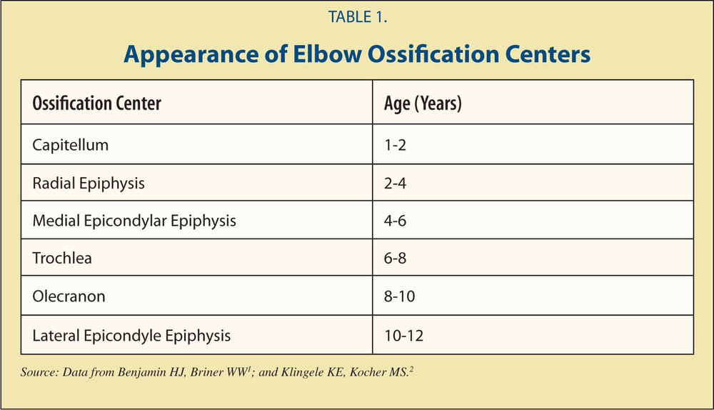
Conference Notes 1-21-2014

Felder     Study Guide

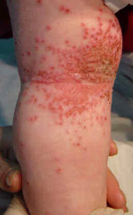
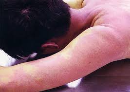
 Pityriasis rosea is possibly due to a viral etiology.  The rash lasts 8-12 weeks.  It is mildly itchy.  It is benign and not contagious.   Girzadas comment: You have to let patients know that the rash will be around for 2-3 months. 


*Pityriasis rosea


Steven Johnson disease is a dermatologic emergency with 5% mortality.  Patients have bullae,  skin blistering, and mucosal lesions.

Elise comments: The diagnosis is TEN if more than 30% of body surface area is involved.   The diagnosis is SJS if less than 30% of body surface is involved.   You have to examine the eyes.  This disease can severely damage the eyes via conjunctival erosion.

 Erythema nodosum is associated with strep infections, TB, and autoimmune diseases.


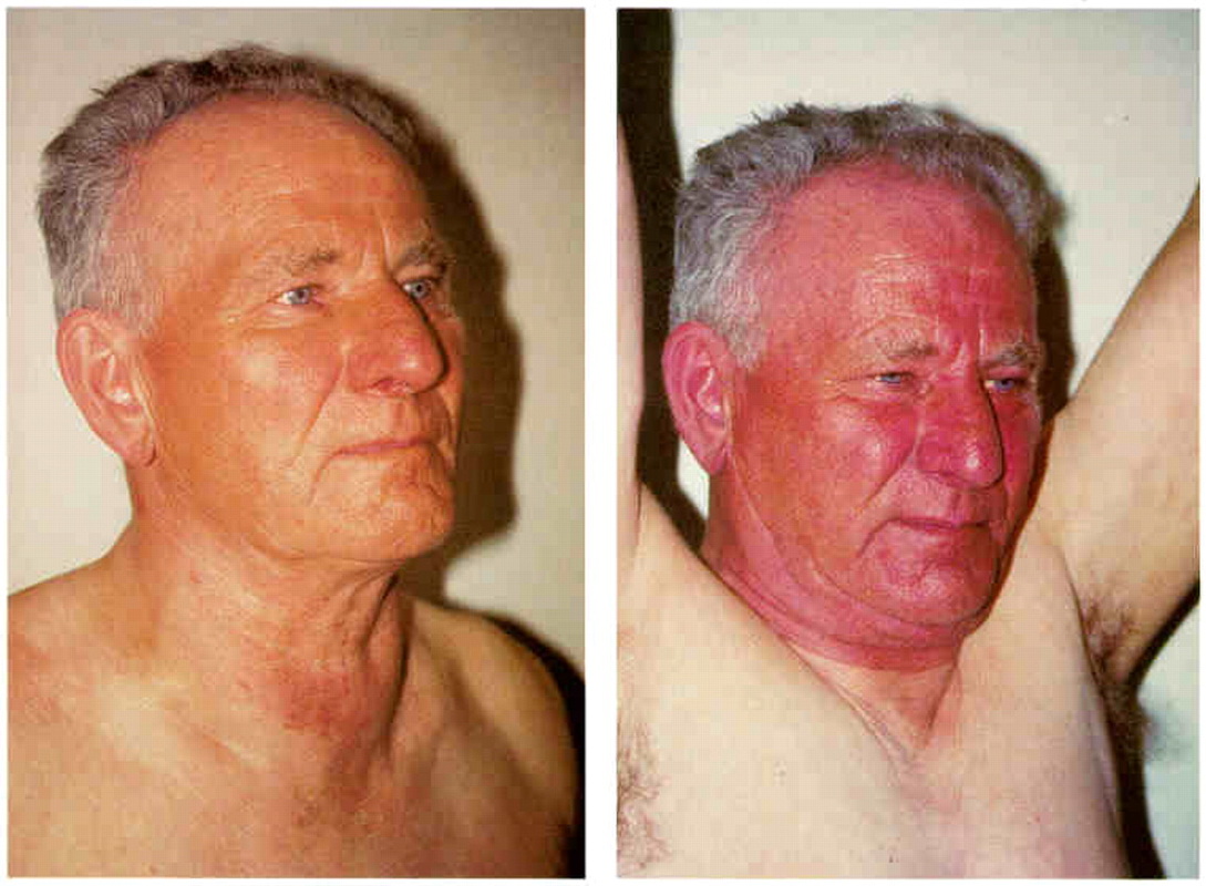
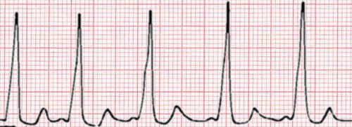
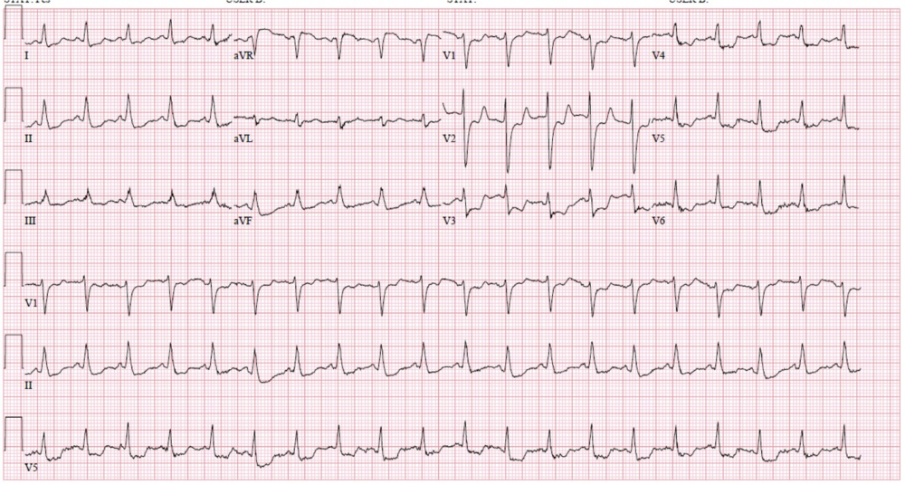
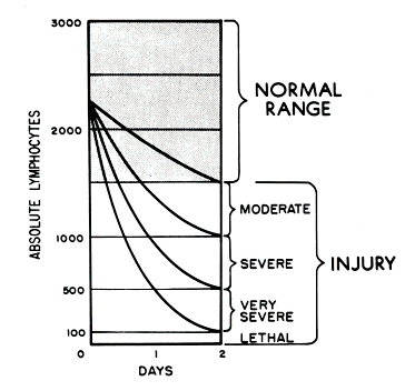
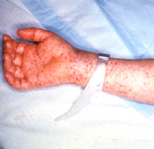
Stage 2 Lyme disease is associated with heart block. 


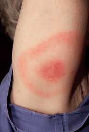
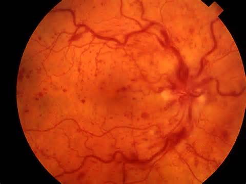
*Erythema chronicum migrans


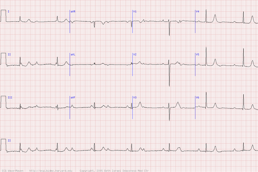
*Heart block associated with lyme disease


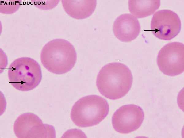
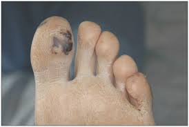
*Rocky mountain spotted fever rash.   Fever can precede rash by a few days. Patients will have headache, nausea and vomiting.


Scombroid poisoning: Results from histidine on the dark meat of fish.  The histidine gets converted to histamine and causes an allergic type reaction.


Fish in the Scombridae family (tuna, mackerel, skipjack, and bonito) are the most common sources of illness. Other fish, such as mahi mahi, bluefish, marlin, and escolar can also cause scombroid fish poisoning.

When these types of fish are not properly refrigerated, bacteria begin to break down the flesh of the fish and histamines are formed. Histamines are heat-resistant; therefore, illness can occur even with fish that is properly canned or cooked. While some contaminated fish will not show any outward signs of spoilage, others will have a bad odor or a “honey-combed” appearance when cooked. Fish that looks spoiled should not be consumed. Histamine levels may be unevenly distributed throughout the fish, and the amount of histamine needed to cause symptoms may vary from person to person. Therefore, people who share a single contaminated portion of fish may show a wide range of symptoms. (Minnesota Department of Health)


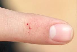
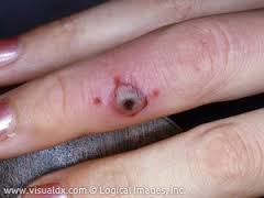
*Janeway lesions are associated with acute endocarditis.


*Silver inhalation or ingestion can cause bluish-grey discoloration of skin.  People who use silver preparations as homeopathy can get this discoloration.  The skin color change is permanent but does not cause any health problem.


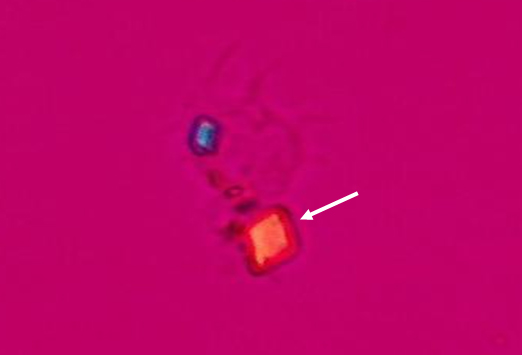
*Disseminated ghonorrhea.   These lesions do not contain bacteria.  You have to culture the cervix or urethra or throat to definitively identify the organism.


CO toxicity is the most common cause of toxicologic morbidity and mortality.   The cherry red skin is uncommon.  Elise comment:  Especially in the winter when people have their furnaces running, think about CO poisoning in patients with headaches,  patients with nausea and vomiting,  and any time two or more family members come to the ED with GI Flu-type symptoms.


*CO toxicity skin changes


Treatment for tinea capitus is griseofulvin for a few weeks.  Topical antifungals don’t work for tinea capitus.  Griseofulvin has some hepatotoxicity, but you don’t need to check LFT’s in healthy kids prior to starting treatment.   Kerion is a more severe version of tinea capitus that is a boggy scalp mass. It also has hair loss.   Don’t incise and drain it.   Treat with oral griseofulvin. 
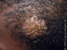
 

*kerion


Berkelhammer         Acute Pancreatitis


Common causes of pancreatitis include: gallstones, etoh, and hyperlipidemia

Many medications can cause pancreatitis including ace inhibitors and valproic acid.

In 1% of patients over 40,  pancreatitits can be caused by pancreatic cancer.


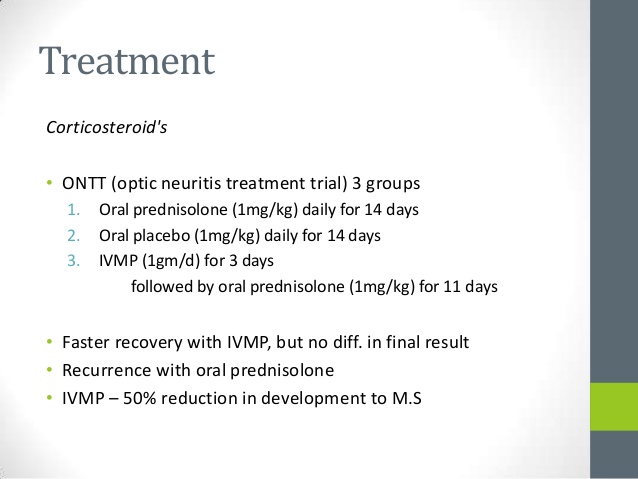
If patients present with pain for 4 days or more, the amylase may have returned to normal.  The lipase remains elevated a few days longer than the amylase.   Dr. Berkelhammer felt that there was no need to order an amylase in addition to a lipase.  He felt amylase just confuses the diagnosis.  Just get a lipase.  


Most pancreatitis is classified as mild with no organ dysfunction.  Patients improve in one week.  You don’t need a CT scan in mild pancreatitis.


Moderate pancreatitis has transient organ failure, exacerbation of comorbid conditions, or pseudocyst/necrosis.


Pancreatitis is classified as severe if the patient has more than 48 hours of organ failure.   This can include renal failure, respiratory failure, hypotension or other organ failure.   High LDH may be an early marker of pancreatic necrosis. 


CT is indicated only for moderate and severe pancreatitis, or if there is a differential diagnosis that needs to be ruled out like perforated ulcer, or cancer.   Girzadas comment: We won’t know by looking at the patient if they have local complication such as pseudocyst or necrosis. So it sound like we should get a scan if the patient has lab evidence of organ dysfunction,  has respiratory failure or has hypotension.  Berkelhammer response: That’s correct.  An IV contrast CT is preferred if  the patient’s renal function is normal.  If the patient has a low GFR, a plain scan will also give useful information.  The choice of whether to use IV contrast or not depends only on the renal function.   IV contrast is not damaging to the pancreas.


Management:   IV fluid bolus followed by maintenance rate of 5 ml/kg/hour for the first 8 hours.    Back off the fluids after the first 24 hours.  Lactated ringers  is preferred over normal saline.

Dilaudid is preferred over morphine due to less ampulla of vater constriction.

Antibiotics are no longer recommended for pancreatitis unless there are signs of sepsis or cholangitis.  Antibiotics have been shown to increase the risk of fungal or other resistant infections.   Antibiotics should be given if you have concerns for cholangitis or patients have signs of ductal dilatation on ultrasound and elevated liver enzymes/bilirubin.  

ERCP is indicated for cholangitis.

Cholecystectomy is indicated 3-5 days after mild gallstone pancreatitis.   If pancreatitis is moderate or severe, cholecystectomy is delayed until pancreatitis is resolved.


Response to Sam Lam’s comments: The level of lipase does not predict severity of pancreatitis.  It is not the BNP of the pancreas.

Sometimes if the serum lipids are very high, the lab cannot run the lipase.  In this situation, you just have to make a clinical diagnosis of pancreatitis and wait for the hyperlipidemia  to resolve.


Parker/Anderson    Oral Boards


Case 1.  56yo male actively seizing.  Patient was an emaciated alcoholic patient.  Patient received high dose benzodiazepines and was rapid sequence intubated.  Patient received IV keppra.  After intubation, a blood sugar was checked and blood sugar was 20.  IV glucose was then given.   Patient then developed torsades while in the ED.  He was given IV magnesium and IV amiodarone.   Torsades resolved with that treatment.   Teaching point was to check blood sugar immediately in a patient that is altered.  The patient was a malnourished alcoholic who was prone to hypoglycemia and hypomagnesemia.

Andrea comment: Think about the Dexi as a vital sign. 

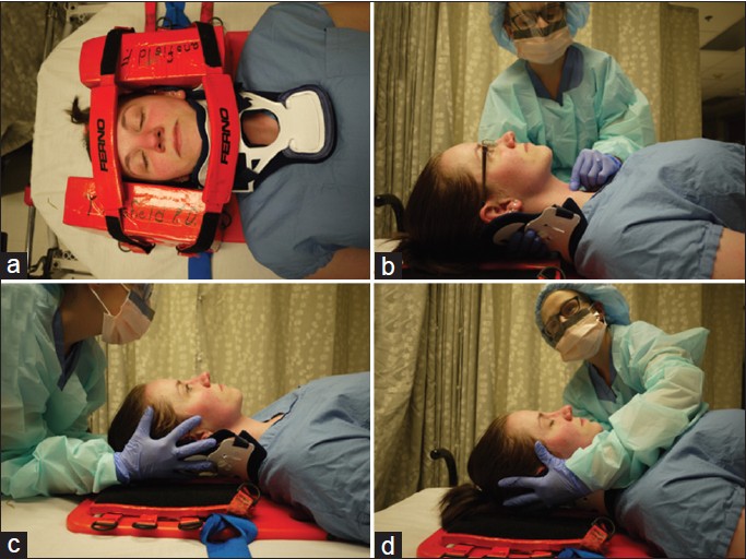
Elise comment: Think about the general description of the patient.  Emaciated alcoholic is at risk for hypoglycemia and hypomagnesemia.   I would also scan his head to evaluate for trauma.  Be sure you know your platelet count and coags prior to performing LP. 


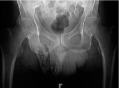
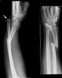
Case2.  82 yo male  brought in by son for constipation.  Patient has dementia.  Vital signs are normal.  Labs show no specific cause of pain.   Son seems perturbed that the work up is taking more time than he would like.   X-ray showed sigmoid volvulous.  Patient is taken for surgical decompression.
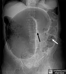
 

*sigmoid volvulus


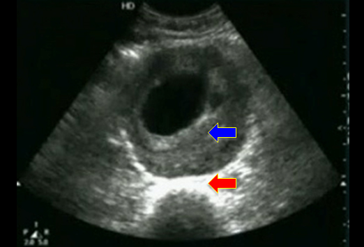
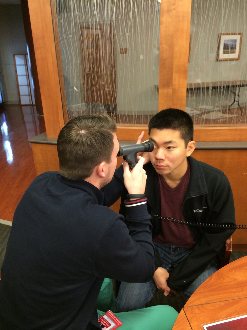
Case3.  65 yo male with eye redness.  Patient has history of rheumatoid arthritis and prior surgery for glaucoma.  Patient receives Humira for rheumatoid arthritis. Patient has HR=102, BP=102/55, T=37.8C.  Eye exam shows hypopion.   Diagnosis was enophthalmitis.   You need to start IV antibiotics. Vanco and Gent are reasonable.  Consider IV anti-fungals as well.   Then consult ophthomology to emergently give intra-vitreal antibiotics.


*Hypopyon


Purnell        M&M


Elderly male presents with abdominal pain.

BP=112/75, HR=108, RR=18, O2 Sat=95%

Patient is in moderate distress due to pain.

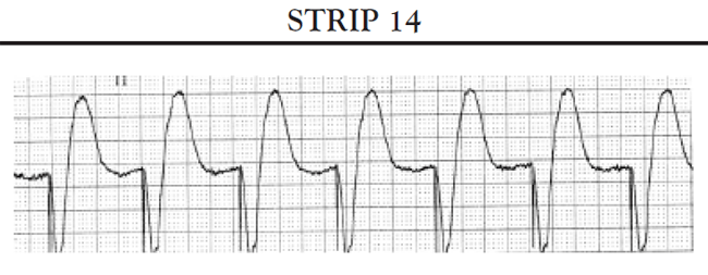
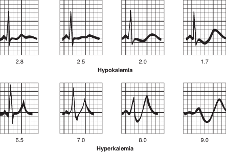
Cardiac exam was irregularly irregular and tachycardic.  EKG shows Afib with RVR.

Abdominal exam had pain out of proportion to exam.  Patient had a large inguinal hernia.

Initial differential diagnosis included mesenteric ischemia, incarcerated hernia, and testicular torsion.

Labs show Lipase of 700 and Lactate is 8.

CT is delayed due crowding.  Patient goes for testicular ultrasound first.  Patient becomes more agitated in ultrasound making CT  difficult and patient is sent back to ED for more pain medication prior to CT.  Patient received IV dilaudid and IV ativan. Patient then becomes somnolent and hypotensive.   He receives full dose IV narcan that brings the patient back to an agitated state.   Still no CT scan done yet and patient is more agitated from pain.

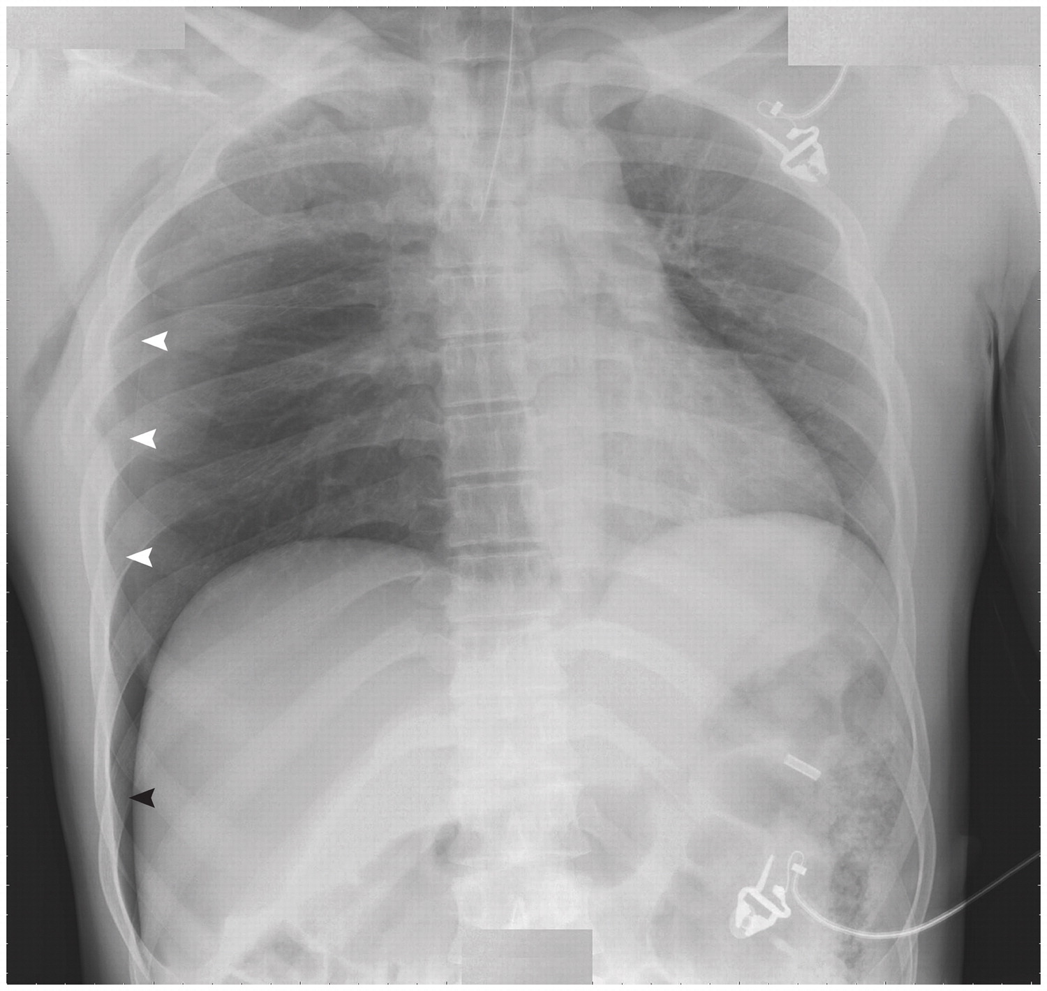
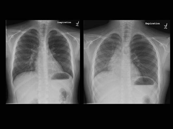
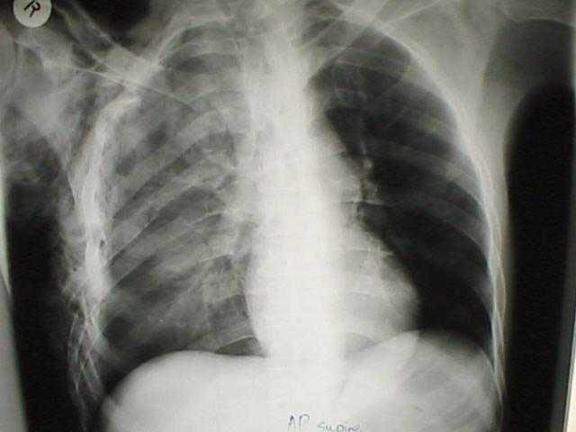
Eventually pain control is opbtained and work up/CT gets done.  CT shows pneumperitoneum and perforated bowel.  Patient was intubated.

Post intubation CXR shows right mainstem intubation.


Teaching points:

You have get the patient to CT in a reasonable time frame.  The physician needs to stay on top of this and follow up with the charge nurse to be sure it is getting done.

Cautiously titrate opioid pain medication and benzos in elderly patients to avoid respiratory depression and hypotension.

In the same way, don’t give full dose narcan to reverse over-sedation.  Tritrate the narcan to the point where the patient is responsive and not hypotensive. 

Check your post-intubation CXR as soon as it is done to be sure of your ET tube position.


Navarette      Safety in the ED   Agitated Patients


60% of all workplace violence occurs in healthcare and social service settings.

A study at Henry Ford identified multiple handguns and knives in the possession of ED patients.


Trauma patients in the ED may be altered from drugs/alcohol  or have a brain injury that could affect their judgment.


Keep a safe distance from patients.  Keep the door of the exam room open.  Stay between the patient and the door so your exit is never blocked. Keep your belongings out of the patient’s  reach. Have a plan of how to get help if you need it.  Trust your gut if you think the patient is showing signs of agitation or potential violence.


Clues to violent behavior: loud speech, pacing, restlessness, suspicious, threatening remarks, responding to internal stimuli, under the influence of drugs or etoh, delirium or psychosis.


Keep your team safe: Stay calm and speak calmly with the patient,  be alert to signs of impending aggression (noted above), don’t threaten the patient or make demands, genuinely listen to the patient when they speak,   try to align the interests of the patient and the medical team, decrease stimuli such as noise and light, give the patient some options like oral benzo’s or food.   Girzadas comment: Studies have shown that food is very effective at lowering the level of agitation/anger. 


Remke comment: Use your security guards to accompany you when going into potentially difficult/dangerous situations.

Lam comment: Make an assessment of the patient’s eyes.  If they are looking at you intensely it may be a sign of impending violence. 


Conference Notes 1-14-2015

McDermott          Study Guide   ENT/Dental

Never re-implant  avulsed primary teeth.

With cribiform plate fractures you have to be alert for CSF leak.   Beta 2 Transferrin is a more specific test for CSF fluid than measuring glucose from fluid draining from the ear or nose.   The residents (Htet and Kennedy) said in the SINI they run a Beta2 Transferrin test on fluid from the nose or ear or even from a swab of the nose or ear to check for CSF leak.

The most common bacterial causes of parotitis or sialoadenitis is staph, strep, and anaerobes.  In nursing home patients you have to use vancomycin to cover MRSA.

Adult epiglottitis usually presents with sore throat and difficulty swallowing.  Adult patients don’t usually present with stridor/drooling.

To fully nerve block the ear you should place an auriculotemporal nerve block.

Elise comment: This block works very well for complex ear lacerations.  Patients get great anesthesia from these blocks.


*Auriculotemporal nerve block


Management of optic neuritis requires IV steroids.  IV steroids improve the time to improvement, have less recurrances of optic neuritis and may prevent the development of MS.  IV steroids do not improve visual outcome over oral steroids.


*Outcomes of IV vs Oral Steroid Trial in Optic Neuritis


Risk factors of posterior nasal packing include cardiac arrest, hypoxia, and necrosis of the colunmella,  Use only as much pressure in the balloons as needed.  All patients with posterior balloon packing need to be admitted.


*Nasal anatomy including columella and philtrum


Ear drops to use when you have a concern for a perforated tm:  Use  cortisporin otic suspension .   Floxin otic solution is another option and is the only FDA approved ear drop for use with perforated TM.    Don’t use any other otic solutions when concerned about a perforated TM  because solutions have lower ph’s than suspensions.     “Otic solution is not the solution”.    Alternatively, you can use ophthalmic preparations of a flouroquinalone.  They are less irritating than otic preparations and in general cheaper. 


Ludwigs’s angina most commonly develops from an infection of the lower molar.


Final Jeapordy Question: Genu 7 Bell’s Palsy is a stroke that looks like Bell’s Palsy but the patient cannot abduct the ipsilateral eye.   Stroke is in the pons.


Katiyar      Critical Care Billing and Coding


Critical care codes have higher RVU’s than other EM codes.

Medicare pays $35.80 per RVU.

A fast track patient generates  about $62 in physician reimbursement.   A Cardiac  patient generates $175.   A Critical Care patient generates $225 in physician reimbursement.


Definition of critical care:  high probability of sudden, clinically significant, or life threatening deterioration requiring the highest level of physician attention.

Simple test of critical care: Does this patient have an illness/injury that if you do nothing, they could die?   If the answer is yes, then it is likely critical care.

  • Some examples of critical care: Being called to the bedside to evaluate opiate overdose with shallow breathing.
  • New onset seizure with fever in a patient with multiple other medications and medical problems. 
  • Evaluation of acute hypotension of any cause.
  • Supraventricular tachycardia requiring Adenosine intervention.
  • Admission for acute renal failure with critical hyperkalemia being managed with or with acute dialysis.
  • Admission for symptomatic hyponatremia requiring 3% NaCl.
  • Admission for pneumonia or COPD exacerbation requiring BiPAP therapy.
  • Admission for large volume gastrointestinal bleeding with tachycardia.
  • Admission for severe DKA requiring an insulin drip.
  • Admission for symptomatic hypertension with headache or chest pain on medication infusion therapy.
  • Admission for rapid atrial fibrillation on continuous Amiodarone infusion.
  • Being called to the bedside to evaluate chest pain with new ST elevation EKG changes.
  • NSTEMI Patient
  • Being called to the bedside to evaluate a patient with acute stroke signs and symptoms. 


There are some diagnoses that you can bill critical care and still discharge the patient:  overdose, anaphylaxis, angioedema, asthma exacerbation.   Be careful of billing critical care with a discharge.  It could trigger an audit.  A medicare audit  is bad news and it covers the last 4 years not just the chart that triggered the audit!


ICU admission is not a requirement for critical care billing.  For example, patients with severe asthma or CHF can be significantly improved by aggressive care in the ED and  then be admitted to tele or the floor.  These patients still qualify for critical care billing.


In addition to documenting the time you spent, you need a  macro stating something to the effect of  “Due to the patients unstable presentation, he required my immediate attention and intervention for  35 minutes    Also document your repeat exams and how the patient was progressing, the interventions you made, and how the patient was responding to treatment.


You cannot bill critical care on a failed resuscitation.


If you are working with a resident, the time you billed critical care has to be your time and not include the resident’s time.    Time spent teaching cannot be counted as critical care.   The same general rules apply if you are working with a midlevel provider.


Critical care time includes documentation time, interpretation of testing, review of prior records, bedside care, re-assessment, discussion with family, and discussion with other physicians.


The time you document as total critical care time is a conservative estimate of the total time you spent managing the critically ill patient.  You don’t need to break down the total time you spent in itemized fashion in your note.  You can do that (5 minutes interpreting ABG, 10 minutes discussion management decisions with patient’s family,  20 minutes at besdside managing hemodynamic instability) but it is not necessary.  


Be very careful to not overbill critical care.  The standard percentage of critical care charts for an EM doc is less than 5%.  On most shifts you can probably bill critical care for one or two patients.   If you are billing critical care over 5%, you are at risk for an audit.


Kalimullah       Critical Care Fellowship Training and Employment


Ejaaz went over the various training routes to becoming a board certified critical care physician.


The IM route has the benefit of  learning to manage many complex, multiply co-morbid patients.  The majority of practicing intensivists are IM trained so politically this track may give you more job opportunities.   The downside is you need 6 months of IM experience, 3 months of which have to be in the ICU prior to supervising IM residents.  That is an RRC requirement for IM training programs.  So you need to do a lot of MICU in residency or at the beginning of fellowship.   The number of EM trained fellows is limited to 25% of total fellow complement in each IM Critical Care program .


The Anesthesia Critical Care route has the benefit of  a common application form and a match.   The curriculum is more tolerant of EM residents.  You don’t have all the IM prerequisites.  You only need 4 months of critical care training during residency.  Anesthesia has a resuscitation-centric philosophy.  There is common interest between EM and Anesthesia with airway management.  You can get TEE training in this track.  You will get a broad clinical exposure including extensive surgical subspecialty exposure.     The downsides are less time spent in the MICU and less exposure to ID/nephrology/pulmonary/cards.  You frequently have to co-manage your patients with the surgeon.

There are only 17 programs nationwide that are set up to take an EM resident as a fellow. Not a lot of spots for EM grads.  Anesthesia grads have shown increasing interest in these spots so there is significant competition.


Surgical Critical Care Route.    Up sides: A lot of experience in trauma and acute surgical illness.   You will likely take care of patients with less co-morbidities than in an MICU.   It can be a springboard for a career in a surgical ICU.  Downsides: Only 6 programs nationwide are set up for EM grads.   The first year of the curriculum is a “supplemental educational program in surgery for emergency physicians”.   This year is basically a year of surgical residency   EM residents are largely taken outside the match.  You will be co-managing patients with surgeons.   Less job opportunities after fellowship because many community hospitals don’t have SICU’s or surgical subspecialty ICU’s.


Employment for EM critical care fellowship trained physicians: 49% do both EM and critical care practice.   25% do critical care only.  21% do EM only.  63% work in academic centers.   If a doc does both EM and critical care in his practice, they tend to work more days than a doc who does only one or the other.  There is something called Crippen’s Law.  This states that by 5 years all dual trained EM/Critical care docs will be practicing 100% in either EM or Critical Care but not both.  This frequently turns out to be the case.


Bolton        Duty and Ethical Dilemma


4 principals of ethics : Autonomy,  beneficience  (work to benefit the patient), nonmaleficence (do not harm) and justice (distribute resources fairly).


Rapid decision tool for ethical questions:

Is there already a standing rule for this situation?  If yes,  follow the rule.  If no then,

Can you buy time?   If yes buy time with medical stabilization.  If no then

be impartial, follow the golden rule.  Your decision should be universalizable.  It should be able to be applied in the same way tomorrow to another patient.  Finally,  you should be able to explain and justify the decision to a lay person.   If you your decision does not follow the golden rule, is not fit to be applied in all situations, or you feel like you can’t justify it to your mom, you should probably rethink your decision.


Case: 16yo female is found to be pregnant and doesn’t want her mom told.    Your response to this request for confidentiality depends on your determination as to whether the patient has capacity to make this decision.    Ask the patient why she doesn’t want this information divulged.  Try to convince the patient to divulge the information to her mom.  If the patient won’t speak to her mom about her pregnancy, then you have to decide if she has capacity to understand the ramifications of her decision.  If you think she does have capacitythan confidentiality is indicated. If you think she does not have adequate capacity, then you need to divulge the pregnancy test result with the patient’s mom and document why you did not feel the patient had capacity to make the decision she did.


Case: Patient with metastatic cancer.  Patient is peri-arrest.  Patient has signed DNR.  Family in the ED says do everything.  The issues here are futility and nonmaleficence.  What the family wants is contrary to the patient’s wishes.   Ask the family why they want the patient to be resuscitated.   The family said that another family member was coming to the hospital and they would want her alive until he got there. 

There was a animated discussion about  whether to intubate the patient to keep her alive until the son arrived.  There was pretty much 50:50 split in the group between those who would or would not.   Ejaaz’s comments:  Autonomy is the most valued ethical principal in the US.  You have no obligation to give futile care.  Some patients will have PTSD after ICU care despite heavy sedation. So sedation while the patient is intubated is not sure to prevent suffering.  Finally there is a slippery slope of having to provide further care beyond intubation to keep the patient alive for those 6 hours.    Discuss with the family that there is a conflict between the patient’s comfort and their desire to keep her alive.  Tell the family that they will have to pick which outcome(prolonged life with some possible suffering vs death with no suffering) they think the patient would value more.   Braden Parker comment: There is also a justice component in this case in which this patient is receiving futile care in the ICU that is keeping another patient out of the ICU.


Ebola issues: Do we as physicians have a duty to take care of an ebola patient?   Because we are a profession we have a responsibility to care for  these patients.   The AMA made a statement in 1983 that physicians have to take care of AIDS patients.  There are some situtions that may take away this responsibility: physicians who are pregnant moms or single parents.  We are not responsible to care for patients without adequate preparation and protection.  However, if there are proper safeguards and protection and knowledge of the disease we have an obligation to care for the disease.   Futility is a factor as well.   There is agreement that we will not intubate or code ebola patients.   Elise comment: It was not a positive time for medicine when in the 1980’s some physicians were refusing to care for AIDS patients.


17yo female with non-hodgkin’s lymphoma. The patient refused chemotherapy.  Chemotherapy for this disease has an 80% success rate.  The patient’s mom supported her daughter’s decision.  A Court forced the teen to undergo chemotherapy.  There was a animated discussion over whether it was appropriate to force the patient to receive chemotherapy over the patient’s and the mom’s desires.


Conference Notes 1-7-2015


Remke/McKean    Oral Boards

Case 1.  2 month old male  presents with excessive crying.  P 176  BP 72/48   RR 28   P/O 99%

Mom notes child has been eating less.   Vomited once.  Child is full term with no health problems.  Exam shows normal male infant except  that patient has a swollen/tender/erythematous scrotum.    Ultrasound shows left sided inguinal hernia.  Bilateral testes have adequate blood flow.   Attempts at ED reduction were unsuccessful.  Surgery was consulted.   Teaching point: Do a complete physical exam on crying infants including the genital area.   Males can have a hair tourniquet, paraphimosis, torsion, and hernia hiding under the diaper.


Case 2. 26 year old female with a chief complaint  that she cannot see with her left eye.  P 108, BP 138/78  RR 22   T 37.  Patient has no headache or eye pain.  No past medical history.   There was a family history of an aunt with a  “clot”.     Patient was on oral contraceptive pills.  CT and MRV imaging of the brain were normal.  Fundocsopic exam shows


*Central Retinal Artery Occlusion


Treatment:  There aren’t any evidence-based therapies.  However you should try digital massage, timolol, and  consult ophthalmology for anterior chamber paracentesis.   Mannitol can also be given. 

Teaching points: oral contraceptives can be a risk factor. 


Case 3.  17yo male with knee injury during a football game.   Vital signs are normal.   Patient noted some transient deformity of his knee immediately following injury.  The deformity spontaneously resolved.    On exam, the knee is unstable.  ABI’s of the injured lower extremity are also borderline abnormal.   Plain x-rays of the injured knee show no abnormality.  CTA of the injured extremity shows normal blood flow. 

Diagnosis was a knee dislocation with a peroneal nerve injury.

Teaching points: Knee dislocations can reduce prior to ED evaluation.  If you identify any neurovascular deficit there is increased risk for other neuro-vascular injury.  The main injury to evaluate for is popliteal artery injury.   


Elise comments: You have to be able to differentiate between central retinal artery occlusion and central retinal vein occlusion.   Once you identify either, you need to move to treat that disease.  Further testing is not going to be fruitful.


*Central retinal Vein occlusion


Carlson    Critical Care Toxicology Updates


Poisoned patients spend less time in the ICU, have lower mortality, and have less mechanical ventilation days when compared to all other ICU patients.


Which toxicology patients should be intubated?

Tox patients don’t necessarily follow the trauma-based rule of GCS < 8 = intubate.  One Dutch study showed the only 20% of toxicology patients with GCS 3-8 required intubation.   If you have to sedate a tox patient to intubate them/keep them intubated consider that maybe you don’t need to intubate them in the first place.   When making an intubation decision, you need to consider the duration of the toxin.  Also consider if there are any reversal agents for the suspected toxin.   


Pulse oximetry can be a useful tool to monitor hypoventilation.  With no supplemental oxygen, the pulse ox will show a drop in O2 saturation that pretty predictably corresponds to a rise in a PCO2.   If you give supplemental O2,  this will blunt the drop in O2 saturation and you can miss hypoventilation.   We then had a lively debate about using supplemental O2 for procedural sedation.  The pro side: you potentially protect against hypoxemia.  The con: It may mask hypoventilation.   The consensus: Give supplemental O2 and use capnometry to pick up hypoventilation.


Hypercapneic respiratory failure is probably the most common reason for intubation of a toxicology patient.   Etoh, benzo’s, opioids, and other sedative hypnotics are common drugs causing hypercapneic respiratory failure.


Hypoxic respiratory failure is less common in toxicology patients but can develop from pulmonary edema or aspiration.


BiPap is less useful for tox patients.   Predictors for Bipap failure include weak cough reflex, excessive secretions, hypercapnea, and agitation.  All of these are common in  tox patients.  Vomiting is another big problem with bipap for tox patients.  The risk of aspiration is very high.   Bottom line: no role for bipap in a tox patient.


Ventilator settings recommended for the toxicology patient:  Assist control,  tidal volume of 6-8cc/kg of IBW (use 8cc/kg if the patient is acidotic/salicylate poisoned), If the patient is acidotic and tachypneic prior to intubation, the respiratory rate you set on the ventilator should equal the patient’s spontaneous respiratory rate prior to intubation.   If the patient has an acid-base critical poisoning they need higher tidal volumes.  Critical acid base poisonings that require higher respiratory rate and tidal volume vent settings include salicylate, barbiturate, and TCA poisoning, and any patient with a critical ph.  If these patients are hypoventilated their acid-base status will rapidly decompensate.


The first hour after intubation is the time when the tox patient will crump. 

In all intubated patients (both tox and non-tox) 25% will have hyotension and 4% will have cardiac arrest in the first hour after intubation.   In tox patients, these numbers are even worse. 


Barounis comment: THAM is buffer that can be given intravenously as  a weight-based drug to counter acidosis. It works like bicarb but unlike bicarb does not get metabolized to CO2 that can cross the blood brain barrier and increase CNS acidosis.


 Barounis      ECMO in Critically Ill Adults


Case:  19yo female post-partum day 2.  Pt had post epidural headaches and failed blood patches.  Patient developed status seizures.   Patient was placed on high dose propofol.    Patient developed propofol infusion syndrome. 


Propofol infusion syndrome:

Propofol infusion syndrome (PRIS) is a rare syndrome which affects patients undergoing long-term treatment with high doses of the anaesthetic and sedative drug propofol. It can lead to cardiac failure, rhabdomyolysis, metabolic acidosis, and renal failure, and is often fatal.[1][2][3] Hyperkalemia, hypertriglyceridemia, and hepatomegaly, proposed to be caused by either "a direct mitochondrial respiratory chain inhibition or impaired mitochondrial fatty acid metabolism" [4] are also key features. It is associated with high doses and long-term use of propofol (> 4 mg/kg/h for more than 24 hours). It occurs more commonly in children, and critically ill patients receiving catecholamines and glucocorticoids are at high risk. Treatment is supportive. Early recognition of the syndrome and discontinuation of the propofol infusion reduces morbidity and mortality. (Wikipedia)


*Propofol infusion syndrome


She develops persistent hyperkalemia and persistent ventricular tachycardia.   Echo shows severe global hypokinesis.  Patient arrests.   She is placed on ECMO and dialysis.  Over several days, the patient’s renal function improves and her cardiac function improved.  She eventually was discharged with full neurologic function and ability to nurse and care for her child.


ECMO is a bridge.  It does not treat any disease  It buys you time to treat a patient.


VA- Full ECMO:  Blood is drained from vena cava and returned to a femoral artery catheter.  In between the EMCO circuit oxygenates and pumps the blood.   There are many options for placing the circuit catheters.  The most common catheter placement is the venous catheter placed in the femoral vein up to the right atrium.   The arterial catheter is placed in the femoral artery.  

With the patient on ECMO the medical team manages the O2 saturation, CO2 removal,  MAP, hematocrit,  and temperature.  MAP is kept around 65.  Hypothermia is induced to increase SVR and improve MAP.   Pressors are also used to maintain blood pressure.  The ECMO pumps do not provide pulsatile flow so the patients have no pulses.  Patients on ECMO are heparinized and at high risk for bleeding.


ECMO  Diagrams


5% risk of intracranial hemorrhage with ECMO.


Dave then made the case for using ECMO in the ED.    ACLS has a low rate of intact neurologic survival.   There are multiple studies showing that  patients who underwent prolonged CPR (30-60+ min) and then reeived ECMO have a good chance of survival with full neurologic recovery.     Dave specifically discussed a study looking at patients with VF/VT arrest who underwent CPR for 30 minutes. ECMO was initiated after 30 minutes of ACLS care.   This study had a 54% percent survival rate of the ECMO patients in which all survivors had good enough neurologic function to live independently at home.   There was a discussion about whether our healthcare system could support the costs of utilizing ECMO routinely in cardiac arrest patients.


HTET      Devices in Human


Case: 50 yo female with tracheostomy tube presents in full arrest.  It was difficult to bag-valve- ventilate patient due to missing inner cannula of tracheostomy.  Another inner cannula was place in the tracheostomy tube and ventilation could be accomplished.  

Tracheostomy tubes:  measurement is based on inner diameter.  A 4.0 Shiley has a 4mm inner cannula.   When placing the tracheostomy tube, have the obturator in place.  When the tracheostomy tube is properly positioned, remove the obturator and insert the inner cannula so that the patient can be ventilated.  


Trach tubes should be changed out about every 3 months.  The inner cannula should be taken out and cleaned every day.


Dislodgment, obstruction, infection, and bleeding are the most common complications of a tracheostomy tube.    Blind replacement is ok >7 days post op.   You can intubate orally if necessary as long as pt has not had total laryngectomy.  If a patient can make some noise orally they likely have not had a total laryngectomy.


Obstruction can be improved with suction, saline irrigation, humidified air and remove/replacement of inner cannula.    If patient is crashing, remove the tracheostomy tube and replace with another tracheostomy tube or endotracheal tube.   Alternatively you can orally intubate if the patient has not had a total laryngectomy. 


Bleeding:  Minor bleeding can be controlled with pressure, packing, or cautery.   Be alert to possibility of a sentinel bleed from a tracheo- innominate arterial fistula.   This occurs between 2 days and 6 weeks after surgery.  The bleeding develops from the innominate artery.    Treat trachea-innominate bleeding by hyper-inflating the trach cuff  in an  attempt to tamponade the bleeding.   If the patient does not have a total laryngectomy you can intubate the patient orally, remove the trach tube, and put your finger in the stoma and compress the innominate artery against the sternum. 


*Compression of trachea-innominate bleeding

Elise comment: If a patient has any bleeding from tracheostomy site between 2 days and 6 weeks after surgery it is a trachea-innominate artery fistula until proven otherwise.  Knowing the timing of the tracheostomy procedure is critical for making this diagnosis early.  You need to admit/observe these patients and consult pulmonary or thoracic surgery.


Air dissection: Air can dissect from trach tube down into the pleural space.  Treat with needle thoracostomy and chest tube.


Tracheal stenosis: This is a delayed complication.  A fibrous scar causes a narrowed trachea.  This can occur a few weeks after de-cannulation.   Treat with head elevation, cool mist, racemic epi nebulizer and steroids.


G-tubes

Clogged: Treat with warm water flush.   Coca cola works well to dissolve the clog.

Deterioration: Yeast implants into wall of feeding tube.  Replace tube.  Ethanol also can be used to sterilize the inner portion of the tube.

Dislodgement: Replace tube if the tube was placed more than 4 weeks ago.   The tube tract should be developed by 4 weeks. 

Faculty discussed G-tube replacement tips:  Sedate with versed or fentanyl.  Intranasal meds for kids work great.  Urojet lidocaine can be used in adults to give some local anesthesia.  Use increasing diameter dilators to dilate the stoma  (cervical dilators, foley tubes, NG tubes).   Get a gastrograffen KUB almost always to ensure proper tube placement.  Harwood felt that if the tube passes easily in to a long term stoma tract and you get gastric contents back you most likely are safe but if you have any doubt at all get a gastrograffen x-ray to verify placement. 

Peristomal leakage: loosen or tighten the external bolster, apply zinc oxide paste.  Can discuss with GI consultant about using larger tube.   The tube size is not a common factor causing peristomal leakage.


Conference Notes 12-17-2014

Gore/Paquette     Oral Boards


Case 1.  Adult male stabbed in the left chest.  Chest tube is placed and patient was intubated.   Patient becomes pulseless.   ED thoracotomy was performed.  Patient was found to have a tense pericardium when the chest was opened.   Incising the pericardium released blood and the patient’s pulses returned.  Diagnosis was pericardial tamponade from a stab wound to the heart.  Once the pericardial sac is opened and the tamponade is released vital signs will often return.  The wound can be stapled or occluded with a finger or foley balloon.  The patient was transported up to surgery. 


*ED thoracotomy


Elise comment: Male patients who need emergent blood transfusion can receive O positive blood.   Only women need O negative blood.   

Girzadas comment: Stab wounds to the chest are the most likely injury in which an ED thoracotomy can save a patient’s life.   These type of injuries can have tamponade without extensive myocardial injury.  If the thoracotomy is done rapidly these patients can survive.


Case 2.  55yo male with severe left flank pain. BP=100/50, P=140.  Patient has history of smoking and HTN.  Bedside U/S shows:


*Bedside U/S

Diagnosis was ruptured abdominal aortic aneurysm.  Patient was transferred to surgery emergently.

Teaching point: If a patient with the history of hypertension develops hypotension you know there is a significant problem. 


Case 3.   3 yo male with seizure and lethargy.  Blood sugar was 10.  History revealed that there was a Christmas party at the home the previous night and the child had ingested left-over ETOH.   The patient’s ETOH level was 180.  Urine toxicology screen was negative.   Critical actions were to correct the blood sugar with dextrose.

Bill Schroeder’s Rule of 50

Age less than one month:  5ml/kg of D10      (5 x 10=50)

Age one month to 2 yrs:     2 ml/kg of D25     (2 X 25=50)

Age over 2 years:                1 ml/kg of D50     (1 X 50= 50)

This patient would receive 1ml/kg of D50 to correct blood sugar.

Also have the crisis worker discuss with patient and consider notifying DCFS for child endangerment.


Iannitelli   Wound Ballistics and ED Management of Gun Shot Wounds

Guest Speaker Darin Crowder US Army Retired Master Sergeant


Case: 30yo male with small caliber/low velocity GSW to left low back.  Patient is alert and talking but BP is 100/50 and heart rate is 130.   Patient has no respiratory distress.   Hypotension improved with IV fluid bolus.  While getting a chest xray, patient becomes bradycardic.   Chest xray shows hemothorax on right and bullet in the right chest.   Patient arrests soon after chest xray.  CPR started, patient was intubated and a right chest tube was placed. Epinepherine given.  Patient had ROSC   Patient made it to OR where he was found to have an IVC/RA junction injury.


Extent of patient injury depends on how much kinetic energy is transferred from the bullet to the tissue.


Shotguns have an effective range of 25 yards.  It is intended to be used to kill birds.  It is an “area” weapon.  Shotgun pellets spread out in a cone fashion across the target area.  There are 300 pellets in a shotgun shell.  The wadding in a shotgun shell can cause injury up to 5-10 yards.


Rifles have a range from 50-6000 yards.  It is designed to be accurate and lethal over long ranges.  


Handguns have a maximum effective range of  around 25 yards.


Maximum effective range does not equal lethality.   Maximum effective range is the range that the shooter can expect to accurately hit a target.   A bullet from a handgun can travel up to a mile and even kill someone at that distance, but the shooter can’t effectively control the trajectory of the bullet for a mile.


Caliber is the diameter of the projectile.  30 caliber is a projectile with a 0.3 inch diameter.    9mm=38caliber=.38inches diameter


There is rifling (grooves) in the barrel of a rifle that causes the bullet to spin which increases the accuracy of the bullet.   Pistols have less rifling in the barrel so there is less accuracy.   With less spin, the bullet will develop yaw  (tilt off of the path of the trajectory) which throws off the accuracy.


Small caliber bullets have a higher initial muzzle velocity  than higher caliber bullets.

Smaller caliber bullets have less powder in the casing.  There is less recoil with small caliber bullets.  Shooters generally prefer smaller caliber bullets so that it is less painful to shoot due to less recoil.


Handguns are typically high velocity(>1100ft/s) rounds. 

Jacketed and non-jacketed bullets:   Jacketed rounds have a lead core with a metallic shell so that the bullet will maintain its integrity as it passes through a target.   Non-jacketed rounds don’t have a metallic covering so the bullet deforms and transfers all its energy into the target.

Hollow point bullets are meant to break apart and deform on contact in order to transfer as much energy into the targeted person.  This is called stopping power.  It is intended to stop a person with a single bullet.


 Damage to the patient depends on how much kinetic energy is transferred from the bullet to the patient. 


  1. Kinetic energy of the bullet at impact is its maximum capacity for injury.  1/2mv2 =kinetic energy
  2. Kinetic energy doesn’t equal energy transferred.  Energy transferred to the target is proportional to low speed (like getting stabbed with a spoon vs. a knife.  If they both penetrate the body the spoon is going to do more damage).  Energy transfer is also greater with yaw, bullet deformity, and larger caliber projectile.
  3. Yaw is when the bullet tilts or tumbles off the direct path it is travelling. 
  4. Permanent cavitation is the damage done by the bullet projectile (crushing/tearing)
  5. Temporary cavitation is the lateral displacement  of tissue around the projectile as it passes through  the body.  Tissue stretches around the path of the bullet.  This causes much tissue damage.


*Yaw and Tumble


Bystanders that are struck by bullets at a distance out of the effective range of a handgun can suffer significant injury because at these distances there is a lot of yaw and tumbling occurring. Thus the bullet hits at a lower speed with yaw and this usually results in a lot of energy transfer to the victim.


Girzadas question:  If low velocity rounds transfer more energy to a targeted victim, why do military weapons  use high velocity rounds.  Darin’s reply:  Milatary weapons/rounds are intended to engage the enemy at a longer distance.  To get accurate shots at longer distances you need higher velocity rounds.  The military has found that in close quarter fighting, high velocity rounds pass through the victim  without stopping them (without much energy transfer).   The military has switched to lower velocity/higher caliber weapons/ammunition for close quarter fighting to stop enemy combatants more effectively.  These low velocity, high caliber rounds impart more energy into the target and have more stopping power.


Doherty comment: It is difficult for front line treating physicians to know whether a gun shot wound is an entry or exit wound.  He advised not making that determination in the medical record because you may have to testify against a forensic pathologist. 


In retrospect, the chest xray had some very telling clues. The bullet had traversed the body from left to right (transmediastinal) and definitely had undergone significant yaw (The bullet was tilted and pointed out of the direction of the bullet path).  The bullet had not deformed so it was jacketed.  Thus the bullet had transferred significant energy to the patient based on yaw and it’s pathway put vital mediastinal structures at risk. 


Doherty comments:  The initial chest xray is a key indicator to where the surgeon should approach the operative repair of gunshot wound injuries.


When looking at xrays the number of wounds plus bullets should be an even number.  If it is not then you need to find another bullet.


Htet       Safety Lecture


85 yo female with atrial fibrillation on warfarin.  Patient presented with abdominal pain.  Patient was sent to CT scan for imaging.  In the CT scanner she fell and hit her head.  She suffered a subdural hemorrhage.  Patient was managed with FEIBA and neurosurgery consultation.


Natalie discussed the Michigan Model of risk reduction which features admitting a  mistake occurred,  quick settlement, and an apology.   This has been shown to reduce the number and cost of claims.

At ACMC, all reported safety events are reviewed/vetted by a patient safety officer and  the PI committee.   A Root Cause Analysis is done if needed.   

Elise comment: Is Advocate moving toward the Michigan model?  Htet response: yes, we still have a way to go but the safety team and risk management are working on it. 


To avoid patient falls in the ED:

  1. High risk patients are identified
  2. Yellow fall-risk wrist bands are applied.
  3. Non-skid socks are used
  4. Patients are assisted to the bathroom


Research has not been able to identify a single risk factor to predict a fall in the next 6 months.  Relative factors related to falls that were identified were depression, foot sores, limited functional mobility, and previous falls.


Lambert    U/S GB, Kidneys, Abdominal Aorta


A nice window to image the gallbladder is through right ribs.   The more traditional window is X -10 or 10cm to the right of the xyphoid.


You want to see the main lobar fissure with the GB in the longitudinal plane.


*Longitudinal view of GB with main lobar fissure seen.


*Gallstones with shadowing


*Wall echo shadow The stones in the gall bladder are close to the anterior wall and shadowing is obscuring the entire gall bladder.


*GB wall thickening is 4mm or more at the anterior wall.   There is also pericholecystic fluid.  A  gallstone also imaged.


*Hydronephosis grading


*Seagull Sign    Celiac Trunk which is the first branch off the abdominal aorta.  Celiac trunk splints into the splenic artery and hepatic artery.


*Abdominal Aortic aneursysm.   Blue arrow is pointing to aneurysm and clot.  Red arrow is pointing to anterior border of spine. 


If you see an older patient with groin pain you should also image the iliac arteries.  Mike has seen 3 iliac artery aneurysms in octogenarians. If you just image down to the bifurcation of the aorta you may miss the iliac artery aneurysm. 


U/S Workshop


Conference Notes 12-10-2014

Permar/Hayward    Study Guide    Heme Onc


Changes in Red cell distribution width (RDW)  precede MCV changes in anemias due to B-12, folate, and iron deficiencies.


Thrombocytopenia is associated with skin/mucosal bleeding  (petechiae, gingival bleeding, epistaxis, heavy menses), also hematuria, and GI bleeding. 

Coagulopathies more commonly present with intracranial bleeding, retroperitoneal bleeding, and hemarthrosis. Girzadas  comment: I have also seen GI bleeding and deep tissue hematomas with coagulopathies.


The clinical finding most commonly associated with ITP is petechiae.   It makes sense because petechiae is in the name of the the disease “ Idiopahtic thrombocytopenic Purpura”.  Acute ITP is more common in kids.  Chronic ITP is more common in adults.


*Liver dysfunction vs DIC   Liver dysfunction will have not much elevation of fibrin degredation products and a negative D-Dimer.


There was a discussion of performing paracentesis in patients with low platelet counts and/or coagulopathy.  Harwood comment: unless the patient is bleeding you don’t need to correct the platelet count or the INR.  Harwood added that every doctor will have a minimal platelet count threshold below which they would be unwilling to do a paracentesis.  His cut off was 50,000.  Elise added that because we use ultrasound to guide paracentesis, there is less risk of puncturing a vessel.  We then discussed doing LP in the setting of low platelets and coagulopathy.  There was consensus that LP could be done if platelets were above 50,000.  No one would do an LP if the INR was prolonged.


Reference: Severe haemorrhage following abdominal paracentesis for ascites in patients with liver disease

  1. I. Pache and
  2. M. Bilodeau


In conclusion, severe haemorrhage after abdominal paracentesis is a rare (0.19%) but potentially lethal complication. We report death following this complication in 0.02% of all paracenteses performed. Most haemorrhages occurred in severely ill patients and were rarely seen in patients with severe thrombocytopenia and/ore elevated INR. All but one occurred in the context of some degree of renal failure. The management of this complication can be conservative. Short-term prognosis is nonetheless poor and seems related to the underlying liver disease. Utility of the systematic prophylactic administration of agents that improve platelet function in patients with renal failure requiring abdominal paracentesis could be assessed prospectively. Unfortunately, this would be difficult to realize because of the very large patient sample required.


Reference:

Brief Report |

Safety of Lumbar Puncture for Children With Acute Lymphoblastic Leukemia and Thrombocytopenia FREE

Scott C. Howard, MD; Amar Gajjar, MD; Raul C. Ribeiro, MD; Gaston K. Rivera, MD; Jeffrey E. Rubnitz, MD, PhD; John T. Sandlund, MD; Patricia L. Harrison, MPH; Alberto de Armendi, MD; Gary V. Dahl, MD; Ching-Hon Pui, MD

[+] Author Affiliations

JAMA. 2000;284(17):2222-2224. doi:10.1001/jama.284.17.222

Conclusions In our study of children undergoing remission induction or consolidation therapy for acute lymphoblastic leukemia, serious complications of LP were not observed, regardless of platelet count. Prophylactic platelet transfusion is not necessary in children with platelet counts higher than 10 × 109/L. Due to the small number of patients in our study with platelet counts of 10 × 109/L or less, conclusions cannot yet be drawn for such patients.


Elise’s Reference: AASLD PRACTICE GUIDELINE

Management of Adult Patients with Ascites

Due to Cirrhosis: Update 2012

Bruce A. Runyon, M.D.

Although some physicians give blood products

(fresh frozen plasma and/or platelets) routinely before

paracentesis in patients with cirrhosis and coagulopathy,

this policy is not data-supported.19,22 Routine

tests of coagulation also do not reflect bleeding risk in

patients with cirrhosis; these patients regularly have

normal global coagulation because of a balanced defi-

ciency of procoagulants and anticoagulants.23 In a survey

of the use of blood products in relation to paracentesis,

50% of approximately 100 hepatologists

attending a conference on coagulopathy in liver disease

indicated that they either never used plasma pre-procedure

or used it only if the INR was >2.5.24 The risks

and costs of prophylactic transfusions may exceed the

benefit. Coagulopathy should preclude paracentesis

only when there is clinically evident hyperfibrinolysis

(three-dimensional ecchymosis/hematoma) or clinically

evident disseminated intravascular coagulation. A

shortened (<120 minute


Bleeding conditions occur in less than 1

per 1,000 patients who require paracentesis. There is

no data-supported cutoff of coagulation parameters

beyond which paracentesis should be avoided.19 In a

study of 1100 large volume paracenteses there were no

hemorrhagic complications despite a) no prophylactic

transfusions, b) platelet counts as low as 19,000 cells/

mm3 (19  106

/L)(54% <50,000) and c) international

normalized ratios for prothrombin time as high

as 8.7 (75% >1.5 and 26.5% >2.0).22


Acute chest syndrome is the leading cause of death in sickle cell disease and most common cause of admission as well.   There was discussion and a search of references on this point  There is controversy whether the most common cause of death is due to acute chest syndrome or infection.  There was agreement that there was overlap between the two etiologies. 

Hydroxyurea has been shown to reduce the frequency and severity of painful crises in children but has not been FDA approved.


*Management of Supratherapuetic INR


Hypercalcemia treatment includes normal saline, calcitonin, zoledronic acid.


*Electrolyte abnormalities on EKG  QT lengthens with hypocalcemia and shortens with hypercalcemia.   T wave varies based on potassium level.


Bonder/Iannitelli        Workshop: ED/EICU  Care of ICU Boarders in the ED


We all worked through the new protocol for the management of ICU Boarders in the ED.  

In situ simulation of the new protocol will occur 1-13-2015.


Campanella    Comprehensive  Stroke Center


We have been successful in getting patients to CT rapidly.

We discussed various strategies to shorten the time from completed CT to administering TPA.


*Strategies for speeding the delivery of TPA   Dr. Campanella discussed many of these strategies.  He had some discomfort of giving TPA in the CT scanner.  He felt that it was more reasonable to use the time of transfer back to the ED to make sure the history and time of onset was as verified as possible and that the CT images were carefully reviewed. 


The eligibility criteria for TPA treatment for stroke in the time period 3-4.5 hours after onset are similar to those for persons treated between 0 and 3 hours, with any one of the

following additional exclusion criteria:

Patients older than 80

Those taking oral anticoagulants regardless of INR

Those with a baseline National Institutes of Health Stroke Scale score > 25

Those with both history of stroke and diabetes.

Findings, on computed tomography of the head, of early infarct involving more than one third of the middle cerebral artery territory


We also disussed the Mr. Clean Trial.  For patients with acute ischemic stroke caused by an  arterial anterior-circulation occlusion, intra-arterial treatment is effective and safe when given within 6 hours of stroke onset, a recent study shows. (Medscape)

This study has only been presented in abstract form.  The manuscript should be out in 2-2015.   Neurologists like it because it backs endovascular therapy for stroke.  There are however some methodological criticisms of the study. 


Beckemeyer     M&M


Parents bring a young child to ED for possible intranasal FB.

Patient has dried brown nasal drainage in nose but with speculum exam, no foreign body was identified.  Full visualization of the nasal passage was difficult due to swelling of the inferior turbinate.

Patient was discharged with ENT referral for the next day.


Child followed up with ENT the next day.  ENT identified a button battery in the nose and by that time the patient had suffered a severe burn to his nasal passage.  He required a nasal stent.


Aspiration of a nasal foreign body is possible but a review of case reports demonstrates that most of the time aspiration occurs with attempted removal of the foreign body.  Only rarely does foreign body aspiration occur spontaneously from a nasal lodging point.  


Button batteries are corrosive to tissue. In addition, they cause necrosis by both electrical discharge and pressure on the tissue.   Button batteries are ubiquitous.  Kids can get them from multiple toys/gadgets in the home.  Musical greeting cards are a big problem because kids can get at the button battery inside quite easily.


Case reports show that perforation and extensive burn from a button battery in the nose can occur within 90 minutes.


Based on this case, it was suggested to ask parents about the possibility of button battery insertion.   Also consider xray imaging of head to identify metallic foreign body in the nasal passage.  Consult ENT  while the patient is in the ED if there is any concern for a button battery in the nose.


Andrea, Elise, and Andrej made the comment that this was a very unusual case, “worst case scenario”.  Based on this case, they all felt that if you have a suspicion for nasal foreign body and you can’t visualize the nasal passage well, get an xray to rule out battery insertion in the nose.  Any non-battery nasal foreign body can wait until they see the ENT specialist to have it removed if you can’t get it out in the ED. 

Harwood comment: What I learned is that if the child has pain in his nose or drainage within a few hours after suspected foreign body insertion I have to strongly consider battery foreign body in the nostril and get an xray.


* Xray image of button battery in the nose.  You have to get this out ASAP.  Call the ENT specialist even in the middle of the night.


Balogun       ABC’s of ED management of Cirrhosis


A= Approach.  Patients present with confusion, lethargy, or bleeding.  DDX is broad including encephalopathy, cerebral edema, intracranial hemorrhage, elevated ammonia level, GI bleeding.   Patients can also present with fever.  You have to consider all potential sources of infection.   Keep spontaneous bacterial peritonitis at the top of your list,  Patients can also complain of edema and ascites.  Patients can have electrolyte abnormalities and coagulopathy.


There was a discussion of the best way to manage the airway in the cirrhotic patient.  General consensus was to do RSI with the patient in head up or RAMP position. Head up position can take some pressure of the diaphragm and improve oxygenation/ventilation.   Use rocuronium instead of succinylcholine.  Succinylcholine can cause hepatic failure in the cirrhotic.  Ketamine can also cause hepatic failure in the cirrhotic.  Most faculty however, didn’t feel there was a huge risk to using ketamine in these patients.


B= Bacterial infections    Cirrhotic patients are immunocompromised.

Spontaneous bacterial peritonitis carries a high mortality.  Diagnose SBP early by performing paracentesis in the ED.   Paracentesis is indicated for patients with fever/pain, prior history of SBP,  ascites and/or GI bleeding and/or encephalopathy.

There was faculty pushback on tapping the bellies of patients with just ascites and encephalopathy or ascites and GI bleed.  Faculty all felt patient had to have some sign pointing to peritonitis beyond ascites like fever, pain, tenderness, or leukocytosis before they would perform paracentesis.

Sole gave this follow up advice via email:  

Who should undergo paracentesis in diagnosing/treating SBP in the ED?
In my lecture, I mentioned that the following scenarios warrants a tap:
- classic sx of fever, abdominal pain/tenderness, ascites
- first time ascites*
- ascites with hx of SBP
- ascites with concomitant GI bleed and/or encephalopathy
Again, these recommendations are based on AASLD (formerly AHA, link to source below) guidelines and is even supported in Tintinalli. During my research, I came across several other sources that support variations of these recommendations. I discussed further with Dr. Raddawi after lecture and his theory regarding performing a paracentecis on first time ascites is that patients who develop ascites for the first time will often times be in decompensated liver failure, worsening their already immunocompromised status. This is also the rationale of ascites with concamitant GI bleed and/or encephalopathy. He does agree, though, that in the presence of normal vitals, no fever, no white count, absence of pain/tenderness, it is a procedure that can be performed inpatient. However, considering the significant mortality associated with SBP, it might be worth it to consider tapping these scenarios in the ED. Incidence of iatrogenic SBP when performed in sterile fashion is <3%


C= Coagulopathy.   Transfuse in platelets are less than 10. INR is the most sensitive sign of worsening liver function.    If INR is prolonged and patient is bleeding give vitamin K and FFP.


C= Circulation.   Cirrhotic patients can be hypotensive.  They are prone to bleeding, volume depletion, infection, and third spacing. Crystalloid replacement is appropriate.  Colloids are indicated only for large volume paracentesis and hepato-renal syndrome.


D= Drug considerations.   Opioids can have prolonged sedation.  Fentanyl is preferred due to its short half life.  

NSAID’s have risk of increased renal toxicity.

Benzodiazepines can have prolonged sedation.  

Zofran should be used in smaller doses.  No more than 8mg in 24 hours.  It can cause hepatotoxicity.

Treat seizures with lorazepam and keppra.  Dilantin can cause hepatic failure.


E= Encephalopathy.   Always get a head CT to evaluate for cerebral edema and intracranial hemorrhage.  Treat with lactulose.  Lactulose traps ammonia and carries it out in the stool.


E Kulstad        ATACH –II Trial


Our ED is a study site for the ATACH-2 trial,  Antihypertensive Treatment of Acute Cerebral Hemorrhage. 


The study goal is to identify if there is benefit to lowering the  BP to less than 140 or less than 180 in patients with acute cerebral hemorrhage.   BP is controlled to these levels for 24 hours.   Patients will be assessed at 30 days for neurologic outcome by blinded neurologists.


Patients are treated with IV nicardipine within 4.5 hours of symptom onset.


There is an iphone/android app for your phone to help you enroll patients in the study.   The app is called ATACH-2.    It has all the enrollment criteria and has a easy to use NIH Stroke Scale.


Conference Notes 12-3-2014

Chastain    Study Guide Endocrine

Treatment of myxedema coma: passive rewarming, vasopressors, steroids,  T4 (4mcg/kg) for thyroid replacement.  Treat hypoglycemia.  Restrict fluids because these patients are usually hyponatremic.

T4 is relatively inactive and gets converted to T3 in the body.  T4 is the most commonly used drug for myxedema coma.  T3 should only be considered in severe myxedema coma.   T3 should be used with caution in the elderly due to it’s potential marked effects.  Michelle has seen STEMI secondary to T3.  Harwood comment: Why would you ever use T3?   Michelle response: In severe myxedema, it may take a long time for the body to convert T4 to T3.  Still avoid its use in the elderly.


When treating thyrotoxicosis, lithium can be used as a substitution for iodine in patients who say they are allergic to iodine.

Elise comment: no one is allergic to iodine.  It is a part of your body metabolism.  Also shellfish allergy does not mean you will be allergic to CT contrast.  It only means you have a slightly higher than normal propensity for an allergic reaction to any drug.

When treating DKA  in pediatric patients, don’t use an insulin bolus.  Be cautious with IV fluids and keep any bolus to no more than 10ml/kg.   Don’t give bicarbonate unless ph is <6.9 and patient is not responding to insulin/IV fluid.

Stress dose steroids for kids is 1-2mg/kg of hydrocortisone.


Michelle and Elise discussed treatment of hypoglycemia in kids. It was agreed that the easiest decision rule was Bill Schroeder’s  Rule of 50.


Age less than one month:  5ml/kg of D10      (5 x 10=50)

Age one month to 2 yrs:     2 ml/kg of D25     (2 X 25=50)

Age over 2 years:                  1 ml/kg of D50     (1 X 50= 50)


Adrenal insufficiency:  Primary adrenal insufficiency means the adrenal gland is not functioning and patients will have hyperkalemia/hypokalemia.  Secondary means there is a problem with the pituitary gland.


*Primary vs Secondary Adrenal Insufficiency


*Diagnostic criteria for Hyperosmolar coma

Harwood comment: In severe hyperosmolar coma , you can sometimes get a lactic acidosis that  will alter the gap and ph from the strict criteria listed.  But it is still hyperosmolar coma and not DKA.

Management of Thyroid Storm:  Propranolol, PTU, Iodine, Hydrocortisone

Consider the mnemonic: PPISS  P=propranolol,  P=PTU,  I=Iodine,  SS=Stress dose Steroids


Joint EM-Peds Conference    Bronchiolitis


New Guidelines published in Pediatrics

Clinicians should not administer albuterol to infants/children with bronchiolitis

Clinicians should not administer epinephrine to infants/children with bronchiolitis

Clinicians should not administer hypertonic saline to infants/children with bronchiolitis in the ED.  Hypertonic can be used occasionally for inpatients as it has been shown to reduce length of stay slightly.

Clinicians should not administer systemic steroids to infants/children with bronchiolitis.

Clinicians may choose not to administer supplemental oxygen if O2 sat exceeds 90%.  Also continuous pulse ox may not be used in infants and children with bronchiolitis.  It is a poor predictor of respiratory distress and can prolong hospital stay.  Spot pulse oximetry checks are preferred.

Chest physiotherapy should also not be used.    Deep suctioning may prolong length of stay and should also be avoided.  Obviously, no antibiotics should be administered for bronchiolitis.

IV fluids can be administered in infants and children who have poor PO intake due to bronchiolitis.

No routine need for viral testing or chest xray.   Dr. Collins states getting a CXR is reasonable in a child with illness more than one week.

Synagis is a monoclonal antibody that can boost the immune system.

Synagis can be used in infants with bronchiolitis who have cardiac disease or chronic lung disease.  It also can be used in premies born before 29weeks.

Audience comment: PCR for RSV can help the clinician rule out a bacterial illness if it is a positive.  Dr. Collins agreed that it is still ok to order PCR’s in atypical cases. 

Beta agonists not indicated unless the patient has had multiple prior wheezing episodes and has a parental history of asthma.   Patients with these characteristics can receive a trial dose of beta agonists.  If they improve, they can get repeated beta agonist therapy.


We will continue to use upper airway suctioning here at ACMC.  No deep suctioning.  

For hydration, IV or NG hydration is acceptable.  


The ACMC experience on the floors showed that  PCR testing had no benefit. 


Elise comment:  In the really sick kids what should we do?   Ahkter response: Heliox, and high flow nasal cannula o2.


Negro/Kerwin        Oral Boards


Case 1.  51yo male with vomiting/diarrhea, weakness.  Patient also has crampy abdominal pain.  Vital signs show tachycardia.  Physical exam reveals ptosis. History reveals that patient ate home-canned peppers.   Diagnosis: Botulism.  Treatment: botulinum anti-toxin, supportive therapy.

 *ptosis with botulism

Case2. 27 yo male with altered mental status. Vital signs unremarkable.  On exam, patient has jaundice. Further history revealed that patient was eating muschrooms while camping on the west coast.  Diagnosis was amanita phalloides mushroom poisoning.  Patients suffering from this toxicity have late onset vomiting (more than 6 hours after ingestion). They can also have liver failure.  Treatment is supportive, manage hypoglycemia, and transfer for liver transplant.  NAC usage has not been shown to be definitely helpful.


*Amanita phalloides mushroom


Case3. 52yo male with vomiting, diarrhea, and weakness. Vital signs show hypotension and tachycardia.   Exam shows abdominal tenderness.  Patient had severe abdominal pain and voluminous stool .  Diagnosis was salmonella poisoning.  Treatment: send stool cultures, fecal leukocytes may suggest a bacterial cause of diarrhea, antibiotics are indicated for severe symptoms/age over 50/immunocompromise.  


Heilicser        Lessons from Katrina and Haiti Response Efforts

Excellent discussion of the difficulties faced by healthcare workers in these disaster settings.


When you are a responder in a disaster, be sure you get dedicated rest time.  IMERT has a rule of 12 hours on and 12 hours off.  If you don’t get rest away from the site you will be come ineffective. 


Harwood question: What are the documentation requirements in a disaster?  Response: Basic patient identifying info and pertinent positives were only documented.  The document stayed with the patient. 


Doctors without Borders is the Gold Standard of Physician Disaster Response Organizations.


You have to prepare yourself psychologically before going to a site like post-earthquake Haiti.  Whatever you expect you will face, it will actually be worse than expected when you get there.  It can be an emotional experience.


You face many ethical quandaries in situations like Haiti.  Who do you resuscitate?  If you resuscitate them how do you continue critical care?  Should you do surgery if you don’t have antibiotics to treat the patient after surgery?   Can you give 10 units of blood to save a single patient when you have other patients who need the blood as well?


In Haiti, placebo medicine with words of encouragement was valid treatment.


Conference Notes 11-26-2014

Febbo        M&M

35 yo female presents with hyperglycemia and left lower abdominal pain.  Vital signs normal except tachycardia to 107.  Physical exam showed left lower quadrant tenderness with no peritonitis.  UA showed a few RBC’s and 1-5 WBC’s . CT Abdomen showed a kidney stone in the left ureter with some obstruction, hydronephrosis, and stranding.  Patient’s pain improved with treatment in the ED and patient was discharged home.

The next day the patient returned with fever and tachycardia (SIRS).  Blood sugar had increased.   Patient was septic due to an infected, obstructed pyelonephritis.  UA on second visit showed high WBC count and bacteria.  Emergent decompression of the urologic tract was performed.

The 5 I’s for identifying the underlying cause of hyperglycemia: Infection, infarction, infant on board, indiscretion with diet or etoh, lack of insulin.

Indications for admission of patient with kidney stone : pain, single kidney, urosepsis, acute renal failure.

Infected hydronephrosis can be a difficult diagnosis.  Lab testing is not always definitive.  Pyuria should be present but can be minimal. Serum WBC is poorly predictive. Urine culture lacks some sensitivity and specificity.

Harwood comment:  The obstructing stone in the ureter can block the downward stream of urine from the infected kidney and result in urine results that underestimate the severity of infection.

There are two options for decompressing the kidney: ureteroscopic removal of stone or IR percutaneous nephrostomy.

Brian went through multiple system issues that contributed to the difficulties with this case including: communication of vital signs from nursing to physician and difficulties with paging the consultants.

Harwood and Elise made the strong point that an infected, obstructed kidney can kill a patient rapidly.   There was a discussion of how you could have picked up this diagnosis on the first visit.   The initial urine study was not helpful.  You have to look at vital sign changes, level of hyperglycemia, and the extent of ct  findings.  Elise and Harwood both stated that they are vigilantly looking for infected, obstructed kidneys with every kidney stone patient they see due to the lethality of this disease.


Sola comment: IR is more likely to drain the kidney during off-hours than GU consultants.

Harwood comment: If you are at a place where GU won’t come in urgently, call IR to treat the problem.  If IR won’t come in then you have to call back GU and demand that they come in.  If neither service will drain the kidney, then transfer them to a hospital that will.  This has to get drained ASAP or the patient will die.   Elise comment:  IR is the better choice for drainage because these sick, potentially unstable patients don’t have to go to the OR.


Parker comment: The hyperglycemia on the first visit was a marker that there was an underlying infection.  We don’t do a great job of identifying why diabetic patients are hyperglycemic.  Emergency physicians must look for an underlying cause.


Harwood and Febbo both made the point that the stoic patient is a marker for serious underlying illness.  Harwood has a low threshold for admitting stoic patients if he is unclear on their diagnosis.


Neal Lyons     Update on Heparin Dosing Protocols


Heparin dosing protocols are based on intensity of heparinization desired.

Reduced intensity: No bolus and starting rate of 12u/kg/hr

Moderate Intensity (Cardiac reasons): 60u/kg bolus (4000u max) followed by 12u/kg/hr  

High Intensity (DVT/PE): 80u/kg bolus (7500u max) followed by 18u/kg/hr  


Beckemeyer       Pattern Recognition


I am just including a limited sampling from this lecture.  There were so many great slides/diagnoses.


FUGU poisoning from eating puffer fish.   This is toxicity from tetrodotoxin.   Supportive ventilation can allow patient to survive.

Symptoms, Treatment, Decontamination

Syndrome Name

  • Puffer fish poisoning
  • Fugu poisoning
  • Tetradon poisoning

Symptoms

  • Tingling,
  • numbness,
  • weakness, lightheadedness
  • limp paralysis leading to dyspnea, cyanosis, cardiac arrhythmia leading to death

Onset of Symptoms

Within 20 minutes to three hours

Rapid diagnostic assay

None available. Sensitive chemical assays for tetrodotoxin have been developed but not approved for use.

Antidote

None available

Supportive Care

Artificial respiration to support breathing


*Supracondylar fractures.  Anterior humoral line should bisect the middle of the capitellum and it doesn’t in the xray on the left.  The xray on the right has a relatively normal anterior humoral line. It still may be alittle anterior suggesting posterior displacement/fracture. Both films have evidence of posterior fat pads which is always abnormal and suggests fracture.


*Perilunate vs. lunate fracture


*Kawasaki’s syndrome


*Kawasaki criteria


*SVC syndrome. Normal on left, plethora and swelling due to SVC on right.  Usually due to a mass in the chest obstructing the SVC.


*WPW rhythm strip.  Note the short pr interval, widened qrs with slurred upstroke


Meyers           The Acutely Painful Joint


Is joint pain due to traumatic injury  or not?   If not, you have to consider infection, crystallopathy, and other things like RA, SLE, viral disease, and lyme disease.


Think septic joint in patients with acute onset pain, fever, immunocompromised patients, those with prosthetic joints and those with rheumatoid arthritis.  Staph is the most common etiology.   Inpatient mortality of septic joint is as high as 15%.   One key exam point is pain with range of motion is 100% sensitive.  If patient has non-painful range of motion they don’t have a septic joint.   There are no absolute contraindications to arthrocentesis.  Relative contraindications include overlying cellulitis and coagulopathy.


Arthrocentesis: Sterile procedure. Prep skin with iodine or chlorhexadine.  Use a 16-18g needle.  Use a bigger syringe for bigger joints.  Smaller syringe for smaller joints.  Larger syringes provide more suction and can accommodate a larger volume of synovial fluid.


For knee arthrocenteses insert your needle in the area of the superior lateral aspect of the patella.  

 John went through arthrocentesis approaches for other joints.  Basically review your approach with a video or text prior to performing the procedure on a patient.

If the synovial fluid cell count is >50,000 this is considered more likely to be a septic joint.  This cutoff is not perfect.  There is significant overlap between septic joint and gout.   There is data that a synovial fluid Lactate >10 is a strong indication of a septic joint.   John tried to get a lactate level run on synovial fluid from taps he performed.  He was unable to get lab or respiratory to run a lactate on synovial fluid here at ACMC.  


Empiric treatment for suspected septic joint is Vancomycin.  You also need to consult orthopedics for surgical washout or serial arthrocentesis Q 12 hours.   Surprisingly, serial arthrocentesis works as well as surgical washout for septic arthritis.


Gout treatment: No NSAID is proven better than the other.  So, use ibuprofen 800mg PO Q8 hours.    If a patient can’t take ibuprofen the you can prescribe colchicine.   Stop both treatments 2 days after symptoms improved.


There are risks to injecting local anesthetics or steroids into a joint.

Harwood comment: I don’t inject local anesthetics or steroids into inflamed joints after I drain them.  Elise I will inject joints if the patient has had prior injections and it helped them.  I don’t do it routinely.  


If you get blood (hemarthrosis) on joint aspiration you have to be alert to the possibility of ligamentous injury, meniscal tear, or fractures of the joint line.


John discussed the orthopedic knee exam.


One nice comparison he noted was the warm-cool-warm  vs.  the  warm-hot-warm  exam.   In general the knee should be cooler than the thigh and lower leg (warm thigh-cool knee-warm lower leg).  In the inflamed or infected joint, the thigh will be warm-knee will be hotter- the lower leg will be warm similar to the thigh.


Harwood comment:  Ultrasound can be very helpful in identifying joint fluid and improving your ability to drain it with arthrocentesis.    My decision points are: Bacteria on synovial fluid analysis, patient comes in and gets started on IV antibiotics.  WBC counts less than 25,000, patient usually goes home.  WBC counts above 25,000 I will usually admit for further evaluation.  

Harwood’s Gout cocktail: 1 dose of colchicine, 1 dose of decadron, and high dose ibuprofen.  I stay away from Indocin due to it’s high side effect profile.


Burns                            Drowning Emergencies


500,000 deaths per year world wide.

Leading cause of death in kids in warm weather states.


Definition: primary respiratory impairment from submersion in a liquid medium.

Process:

There is voluntary breath holding for about a minute

When water gets in the throat there is reflex laryngospasm

Laryngospasm eventually relaxes

Active breathing of liquid

Hypoxemia

Lung injury

Neurologic damage


Management:

Pre hospital: ASAP give 2 rescue breaths then initiate ACLS

Provide O2

Cspine immobilization indicated only for significant blunt trauma

No Heimlich maneuver.


ED: Intubate or provide O2,  Give PEEP

ABC’s

Perform nuero exam

Remove wet clothing

Order CBC, EKG, BMP, Utox, and ABG

CXR but it may not be predictive

CT head/cspine for blunt trauma

NG tube

Albuterol med neb treatments may be helpful

Ventilate with low TV and High PEEP

Arrange broncoscopy for sand or particulate matter in airway

Give volume if needed

Rewarm

Consider a seizure

No need for steroids or prophylactic antibiotics.


Prognosis: Markers for poor prognosis include longer submersion time, longer resuscitation time, warmer water, prolonged time to life support, GCS<5, continued CPR in ED.

Resuscitation should go on for at least for 30-60minutes.  If it was a cold water drowning you should attempt resuscitation to at least 60 minutes.  If the patient is hypothermic at one hour you need to rewarm the patient actively prior to pronouncing the patient.

Be very hesitant to pronounce drowning victims in the field.  All drowning victims even of unknown duration should get a trial of resuscitation.


Elise comment: There is a big difference between kids and adults who drown in cold water. Kids cool rapidly and that better protects their brains and they do better than adults whose bodies/brains cool more slowly.  Nevertheless,  you have to take each patient on a case by case basis.


Harwood comment: I would do a CXR on all patients and observe them.  If there is any abnormal CXR findings, I will admit the patient.


Schroeder/Negro    Wellness and Nutrition


Traditional Low Fat diet philosophy: Avoid fat because it has the highest caloric density.   In the 1950’s Ancel Keys presented  research that touted the benefits of a low fat diet.  This philosophy was incorporated into government guidelines.  The food pyramid then became based on carbohydrates.  The low fat diet has been the largest social experiment in history.  In the US, a low fat diet has resulted in obesity.  Sugar activates dopamine reecptors in our brain similar to cocaine/heroin.   It also upregulates our hunger hormone receptors.  Consequently with a low fat high carb diet you tend to eat more. 


Our body is a finely tuned machine with a delicate balance of hunger and satiety hormones.  Sugar messes up this balance.


If we change to eating more fat do we increase our risk of high cholesterol?  There has never been any data showing increased cholesterol results in increased CV disease or mortality.    If you want to focus on your lipids, lower your triglycerides and increase your HDL.  The best way to do this is eat les sugars and live an active lifestyle.


Eat natural foods and avoid processed foods.  Wild salmon, flaxseed, chia seeds, grass fed beef and bison and enriched eggs all are good sources of Omega-3 fats.

Decrease your omega 6 intake by watching your soy oil and vegetable oil intake.


Good fats to eat are medium chain triglycerides (coconut) and monounsaturated fats (olives, avocado, dark chocolate).   When choosing dark chocolate, the darker the better and it should be in the un-dutched or non-alkalinized format.  Williiamson comment: Dark chocolate should be at least 70% cocoa to get health benefits.  


It is better to eat the whole food rather than just the oil or extract.  For example eat the whole coconut, not just the oil.

Beets lower your blood pressure.

Potatoes and corn are not vegetables. They are grains.


Things that are not good for us: Less sleep,  inadequate fluid intake, yoyo dieting.


Being active throughout the day is better than exercising.  Exercising is just another way of depriving yourself of calories. Muscle building is the important for overall health.  Muscle mass burns more calories and tends to keep you leaner.


Eat when you are hungry, eat enough protein, avoid simple sugars, eat vegetables with every meal throughout the day.


The less processed the food the better.


Conference Notes 11-19-2014

Kmetuk/Katiyar     Oral Boards


Case1.   8yo male very uncomfortable on cart due to pain.  On exam patient has tender abdomen with palpable muscle spasms.  Patient also has priapism and painful muscle spasms of extremities.  Evidence of a spider bite was also noted on the patient’s skin. Further history from grandmom  brings out the fact that the child was playing in the woodshed prior to the onset of pain.   Diagnosis was black widow spider bite.  Patient was treated with narcotic pain medication and black widow spider antivenin.  Also benzodiazepines are effective for muscle cramping. Patients should also receive local wound care and tetanus prophylaxis. Intravenous calcium is no longer recommended. 


*Black widow spider


*Black widow spider bite


Teaching point is that BW spider bite victims will present more commonly with  muscle cramps than with a complaint of a spider bite.  Abdominal muscle cramps are the classic presentation.  You can also get vital sign abnormalities and uncommonly priapism.


Case 2.  Elderly woman was found unresponsive in her bed.  Her apartment was noted to be very hot.   Patient’s temperature was 40.5 C.  CPK was very elevated.  Diagnosis was environmental heat stroke with associated rhabdomyolysis.   Critical action was to rapidly cool the patient.   Also, hydration was indicated to treat rhabdomyolysis.  The emergency physician should also rule out sepsis and toxins as the cause of altered mental status and fever.   Rapid evaporative cooling can be achieved by spraying room temperature water on a disrobed patient and directing fans on patient.   If you have a therapeutic hypothermia device such as the arctic sun device that is another effective means of rapidly cooling the patient.


Case 3. 32 yo male with respiratory distress and rash after being stung by  bees.  Patient was diagnosed with anaphylaxis and was resuscitated with epinephrine, diphenhydramine, steroids and cricothyrotomy.


*Anaphylaxis criteria


Harwood comments: The arctic sun device was very effecting in cooling a hyperthermic patient we managed  last week.   Tylenol will not be effective in lowering the temperature in a hyperthermic patient.   You will have to initiate active cooling measures like cooling device or evaporative cooling.


Burt   Safety Lecture


Current clinical practice managing critically ill patients in the ED is not optimal.   If patients have a delay of 6 hours to get an ICU bed it increases hospital length of stay and increases suboptimal outcomes.


EICU (critical care telemedicine) has been shown to improve mortality, decrease ICU complications, and shorten ICU length of stay.


We are going to trial a new process of caring for ICU boarders in the ED.  These patients will have increased nurse staffing to the level of  a typical ICU (2:1 nurse to patient ratio).  ED physicians and EICU intensivists will be following the patients.   Orders will be entered into Allegra and mesh with the in-hospital system while the patient is still in the ED.  Communication will be improved/increased between the ED/ICU/EICU.  There will be a 5 way phone call between the ED physician/ED nurse, ICU physician/ICU nurse, and EICU physician.


Febbo    Society Recommendations for EM


*Choosing wisely campaign for EM


Oral Rehydration is preferred over IV Fluids in vomiting children.

Give the child 5 ml orally  every 2 minutes.  Pedialyte is the optimal solution.  Goal is 100ml/kg over 4 hours.   Can pretreat with Zofran if the child is vomiting.  Vomiting is not a contraindication to oral rehydration.


Elise comment:  If you are trying to rehydrate a pediatric patient you have to give them more than a popsicle.


Cirrone comment:  Dr. Sawlani said the top diagnoses missed when managing vomiting kids was pyelonephritis, lower lobe pneumonia and brain tumor.   So keep those in mind when caring for a child with vomiting and dehydration.


Avoid CT head in patients with syncope.  They are unlikely to provide useful information for patients with syncope.


Avoid CT Chest for PE in patients with negative PERC rule and in those with a negative D-Dimer and Wells score less than 4.  The risk in these patients is low enough that radiation risk exceeds the diagnostic yield.  Harwood comment: You will miss some PE’s with this approach but it still is the right way to go.


Avoid lumbar spine imaging in atraumatic low back pain unless you have suspicion for serious underlying process.


Avoid antibiotics for treating sinusitis.  Consider decongestants and nasal steroids.   If you give antibiotics Augmentin should be your first line.  Doxycycline and flouroquinolones are alternatives.   Harwood comment:  I recommend treatment with antbiotics for patients who have been ill for more than a week.  In the first week of a sinusitis-type illness, I agree that antibiotics are not helpful


Avoid CT abdomen/pelvis for patients under age 50yo with history of renal colic and a presentation consistent with prior stone.   You can use ultrasound in these patients to identify hydronephrosis.   You need to also check a urine for infection.  Probably should also check a creatinine.


Carefully consider obtaining a urine culture in older patients because there is a 50% rate of asymptomatic bactiuria.  Don’t get a UA/culture unless they have symptoms suggesting a UTI.  Make sure you are considering other causes of  the patient’s symptoms and you aren’t just latching on to a false positive UA result.


Don’t rely on antihistamines to treat severe allergic reactions.  You have to give epinephrine.  Girzadas comment:  I sometimes see hesitancy to give epinephrine.  If the patient has criteria (see chart above)  for anaphylaxis, give epinepherine subQ.  After that you can give Benadryl, H2 blockers, and steroids.


Topical antibiotics are not indicated for surgical wounds.   Harwwood comment: This really means don’t use neomycin on wounds.  There is a high rate of contact dermatitis due to neomycin.  When patients apply neomycin or triple antibiotic containing neomycin they get an erythematous skin reaction. This reaction is bothersome to the patient and can look like a wound infection.


Consider not prescribing antibiotics for otitis media in kids between 2-12 years old if they don’t have severe pain and don’t have high fever (>39C)


American Academy of Ophthalmology recommends no antibiotics for adenoviral conjunctivitis.  All faculty agreed that this is a tough sell to parents and schools.   Harwood comment: Not treating with antibiotics and having patient follow up with ophthalmology is probably a much more expensive and inconvenient option than just giving topical antibiotic.


No need to give FFP or Platelets to patients prior to paracentesis. 


Avoid NSAID’s in patients with HTN, CHF,  or CKD of any cause.


Carlson     Antidiabetic and Hypoglycemic Agents


Consider hypoglycemia in every patient with altered mental status and/or any focal neurologic finding.  Andrea discussed a patient who presented altered mental status and tonic eye deviation.  The presumptive diagnosis was intracranial hemorrhage and the patient went to CT.  The CT was normal but the glucose was 18.   All faculty agreed the hypoglycemia will fool you. 


If you suspect intentional insulin overdose:  Examine skin for boggy and/or hemorrhagic area. High doses of subQ insulin will have a significant skin reaction.   Check Insulin C Peptide level.  Exogenous snsulin overdoses have high insulin levels and have low C-peptide levels.  Endogenous insulin contains c-peptide.  Exogenous insulin does not have c-peptide.


Intentional insulin overdose:  23 hour OBS if the patient is hypoglycemic at any point.


Sulfonylureas stimulate endogenous insulin release.  C-peptide levels will be elevated because it is stimulating endogenous insulin release.  A single tablet can make a pediatric patient hypoglycemic.   Basic management recommendations for sulfonylurea overdoses: 23 hour observation, IV octreotide for recurrent hypoglycemia, IV dextrose for hypoglycemia but no prophylactic IV dextrose in the normoglycemic patient, early feeding, regular accuchecks, and observation of mental status.   Meglitinide overdoses are managed similarly to sulfonylureas.


If patients need D25 infusions to maintain normal blood sugar, you will need to place a central line.


Octreotide is indicated for sulfonylurea overdoses if the patient drops their sugar a second time after initial dextrose therapy.  Andrea said you could make a case to give octreotide after the first episode of  hypoglycemia but she felt most toxicologists would wait for a second episode of hypoglycemia.  With have fewer episodes of hypoglycemia and lower glucose requirements.  It is given subQ or slow IV infusion.


Metformin inhibits gluconeogenesis and enhances peripheral glucose uptake.   Hypoglycemia is rare with metformin overdose.  Overdose with metformin can result in lactic acidosis (MALA).  Metformin in high doses is a cellular poison and disrupts  aerobic metabolism.  In high doses it acts as a weak cyanide-like substance.   If a patient develops lactic acidosis, dialysis is indicated.


Permar       Intro to Reading Chest Xrays


CXR Interpretation Mnemonic:  Drs.  ABCD

D= Data of patient (name, age, MR#)

R= Rotation/inspiration/exposure

S= Soft tissues and bones

A= Air, airway

B= Breathing (lung)

C= Circulation (cardiomediastinum)

D= Diaphragm

E= Extras (central line, NG tubes, ET tube, pacer, etc)


Trale took us through multiple chest xray examples.


*Deep sulcus sign for  Pneumothorax 


*Free air under the diaphragm


Beckemeyer       Patient Satisfaction


On the interview trail Jen has found that employers are asking if our program has formal patient satisfaction training.  Understanding of patient satisfaction principles is a big interest of employers.


Press Ganey surveys query patients about the physician: Friendliness, explanation of the diagnosis, shared decision making, clear instructions, clarity of medication information, amount of time the caregiver spent with you, the physician’s attention to the patient’s comfort, and the patient’s  confidence in the provider.


Elise comment:  Everything on this survey is about communication. It is not about how smart you are or whether you made the correct diagnosis.  All of us can employ communication strategies to do better on the survey.


Easy things:  Patient privacy, food, water, work note, school note, and clean environment.

If you have to take a phone call during your H&P, apologize to the patient for the interruption, tell them the call will be brief and step out of the room to take the call.


Treat pain early and aggressively.


Body language:  It is best that the physician sits.  If you must stand, avoid crossing your arms as that seems to indicate you are closed off or hostile.  Make eye contact.  Introduce yourself to everyone in the room.  Avoid interrupting the patient when they are speaking .  Check on the patient frequently.


Elise comment: If patients are using medical terms, ask them if they are in the medical profession.  If they are, their knowledge helps you a lot.   Be sure to smile.  Smiling is a simple strategy that is extremely effective.

Kelly comment: If you are the second doc to see the patient in the ED, try to preface your conversation with the patient that you spoke with the first doctor and you just want to go over the key points of the history.  You want to have the patient understand that you and the other physician discussed their case and management.  You don’t want the patient to feel they are starting from scratch all over again with another doc.


Demonstrate empathy.  Try to find out what patients are most concerned about.


Navarette comment: Apologize to the patient for their wait and thank them for waiting.   Harwood comment: Apologizing to patients for their wait time or any other issue that has impacted their visit is a very effective strategy.


Give the patient a plan.  Let them know what you are thinking and what the expected course of the ED visit will be.  This really helps the patient understand what is going on while they are in the ED.


Conclude the visit with discharge instructions and thank them.  Make sure they know all the testing and critical thinking that you did for them at this visit. You want them to understand that you put in a lot of attention and thought to their care.


Christine comment: Doing these things make your patients happier and in turn make you happier and make the shift better.   There is a definite payoff to the physician for paying attention to patient satisfaction.  

Harwood comment: It is ok to negotiate with patients about what tests they want and how long they are willing to wait for a specific test.  It gets back to shared decision making.


Conference Notes 11-12-2014

Knight/Gupta                Oral Boards


Case 1.  28yo female, 38 weeks pregnant.   She is complaining of shortness of breath with exertion.  She also notes lower extremity edema.   Pulse ox is 88% on RA.  Lung exam demonstrates bi basilar crackles.   CXR shows cardiomegaly and pulmonary edema.  To complicate the situation, the patient is intent on signing out AMA.  Diagnosis is peri-partum cardiomyopathy.  Critical actions: Monitor fetus, treat CHF with O2 and diuresis and consult cardiology, consider preeclampsia.   Convince the patient to stay in the hospital.    Elise comment: You can’t keep this patient against her will. You can’t forcibly restrain her.  If she has decisional capacity she will have to eventually be allowed to leave.  This is a high risk situation.


Case 2.  9yo male with left  elbow injury.  Patient fell from a slide at school.   Patient has swelling of left elbow/forearm region.   Xray shows a montagia fracture.  Critical actions: manage pain, diagnose montaggia fracture, discuss timing of surgery with orthopedics.


*montaggia fracture (fx of prox third of ulna and dislocation of radial head)  Note that radial head does not line up with capitellum


Girzadas comment:  The key to montaggia and galeazzi fractures are that they need ORIF.   If you are not clear to the orthopedist when taking the oral boards as to the diagnosis, they will tell you to splint the patient and have her follow up.  This will cause you to fail the case.


Case 3.   27 yo female presents with vomiting and diarrhea after eating at a restaurant.  HR-124, BP=129/88. Rectal temp is 37.1C.  On further history, patient also notes a headache and photophobia.  Patient has neck stiffness on exam.   CT head shows SAH. 


*SAH


Elise comment: Don’t get sucked into the diagnosis of gastroenteritis based on the history of vomiting and diarrhea obtained in triage.   Think through other differential diagnoses.   Don’t write gastroenteritis as the diagnosis on the chart.  Better to say vomiting and diarrhea.  With careful history this patient did not have diarrhea actually.  She actually had headache and vomiting and the triage note was misleading. 


Harwood comment: With the oral boards you have to move through the cases quickly.  Use the images /stimuli to key you into the diagnosis.


Parker       Traumatic Brain Injury


 35 yo male found down in an alley.  Airway was OK.  GCS was 10.  Patient had frontal hematomas.  GSW to the occiput.  Presumed mechanism was that patient was shot in the back of the head and fell forward.   CT showed cerebellar bleed and edema due to the GSW.  Patient also had midline shift on higher cuts on the CT.  Incidentally, patient had a retrobulbar hematoma of the left eye.   Patient was intubated and taken to the OR for decompression.   Patient deteriorated in the ICU and eventually was pronounced brain dead. 


*retrobulbar hematoma

ED priorities for the management of TBI: Prevent secondary insult to the brain by avoiding hypotension and hypoxia.   A single occurrence of hypoxia or hypotension results in a 150% increase in mortality.  So, intubate early.  Use fluids and pressors as needed to maintain SBP.  Mannitol can be effective if the patient is at risk of herniation.  Prophylactic hypothermia is not currently recommended.  20% of patients with severe TBI will have a dvt/pe.  Use SCD’s and consider LMWH to prevent dvt/pe.    If the ICP >20 treatment to lower ICP should be initiated.   Keep cerebral perfusion pressures between 50-70.    Prophylactic anticonvulsants can decrease the rate of seizures in the first 7 days.  It does not help decrease the rate of seizures after 7 days.   Hyperventilation can be used cautiously for patients with significantly elevated ICP and at risk of herniation.   Steroids are contraindicated for TBI.  This is the one level 1 recommendation for TBI, no steroids.


Braden recommended the Brain Trauma Foundation guidelines for management of head trauma.    https://www.braintrauma.org/coma-guidelines/searchable-guidelines/


When consulting neurosurgery for TBI, be able to relay the history of the injury, any use of anticoagulants, CT findings, mental status, and the trajectory of the patient’s mental status.  The neurosurgeon needs this info to make a decision of whether to come to the hospital or not.


Elise comment:  Be alert to new evidence coming out about therapeutic hypothermia for TBI.  There are some multi-center studies that will be coming out soon and possibly support new recommendations for the management for traumatic brain injuries.

Christine comment: Remember that Coumadin isn’t the only blood thinner that patients can be on these days.  You may have to carefully ask patients about what medications they are on to not miss other blood thinners like rivaroxaban or clopidogrel for example.


Difficult Conversations  Workshop


Michigan model:  Medical Center Physicians/Administrators apologize to the patient and their families and explore how things went wrong.  They will explain to the injured parties what happened.  They vigorously defend the case when they feel they are right.   Litigation is viewed as a last resort.   Michigan has differences in malpractice law from Illinois that may limit implementation here.


We broke out in to small group simulations covering:

Raising a concern with family for pediatric abuse

Death notification of an adult

Discussion of end of life choices for critically ill/poor prognosis patient

Disclosure of procedural complications

Disclosure of medical error

Death notification of child


General guidelines:

Meet with family as soon as possible.

Have the discussion in an area that is quiet and private.

Optimize your appearance prior to meeting with family (clean scrubs, lab coat).

Have plan in your head and take time to prepare what you are going to say before you get in the room.

Ask the family what they know and what their understanding of the situation is before you give information.

Provide the information as clearly as possible.  If the patient died, you have to use the words dead or died.   

There is controversy about offering an apology.

Give the family time to express their thoughts/feelings.

Express compassion for them.  Offer support going forward. 

Answer any questions.

Provide guidance on next steps.


Conference Notes 11-5-2014

 Permar           M&M

9 day old female born at 38 weeks without complication. Patient presents with jaundice, decreased PO intake, no fever.   T=37.2,  HR=113,  RR=26, BP=81/52, SPO2=100%

Patient noted to be lethargic.  Mucosa was somewhat dry.  Bilirubin level was ordered by the resident.

Attending saw the patient and advised that a septic workup should be done.

 When labs come back, the total bili was 28!  LP was performed and showed CSF fluid that was yellow tinged due to elevated bilirubin.

The Pediatric service advised PICU admission for management of the bilirubin level and possible exchange transfusion.

 Elevated bilirubin develops from increased fetal HGB breakdown.  Neonatal hepatic function is slow compared to older infants and adults. By 14 days of age, most neonates have reached adult level hepatic function and in most infants neonatal jaundice is resolved by that time.


*Bilirubin Metabolism (Colletti EM Clinic Nov. 2007)


Causes of indirect (unconjugated) hyperbilirubinemia: physiologic jaundice, ABO incompatability, sepsis, cephalohematoma, enzyme deficits.   Breast milk jaundice is the persistence of physiologic jaundice beyond one week. Human milk promotes bilirubin absorption in the gut.  Breast feeding failure jaundice results from lactation failure and inadequate fluid intake.  Hypovolemia results in elevated bili levels and increased enterophepatic reabsorption.


The problem: Bilrubin crosses the blood brain barrier and can cause damage to the basal ganglia, hippocampus, and cerebellum, and some cranial nerves.     BIND=B=bilirubin I=Induced N=Neurologic  D=Dysfunction.  Patients with BIND can have hypotonia/hypertonia,  decreased alertness, poor feeding and develop long term cognitive deficits.


Danger signs in the Jaundiced Neonate

1. Family history of significant hemolytic disease

2. Vomiting

3. Lethargy

4. Poor feeding

5. Fever

6. Onset of jaundice after the third day of life

7. High-pitched cry

8. Dark urine

9. Light stools


Galactosemia presents as feeding intolerance manifested by persistent vomiting, an

enlarged liver, seizures, and lethargy.  Ill appearing patients will likely be acidotic.  Newborn screening should pick this up, but there can be false negatives. 


In a well-appearing, afebrile infant with unconjugated

hyperbilirubinemia and normal hemoglobin, no further tests are needed.


Conjugated hyperbilirubinemia is concerning because, although it is nontoxic,

it is a marker for serious underlying disease. Associated findings of conjugated

hyperbilirubinemia are pale acholic stools, dark urine, or the presence

of bilirubin in the urine.  (Colletti EM Clinic Nov. 2007)


The mainstay of treatment for hyperbilirubinemia is phototherapy.  Modalities beyond phototherapy include IVIG and exchange transfusion.  Exchange transfusion has the usual risks associated with transfusions.

Take home lessons:

Be alert for the sick neonate

Consider sepsis in every jaundiced neonate

Be alert for delays in how your work up was going.

Get phototherapy started as soon as possible after identifying a bilirubin >18.

In severely high bilirubin levels (>25) consider IVIG and/or exchange transfusion.


Harwood comment: What was the discharge diagnosis?  Trale: Breast feeding failure jaundice.   Elise comment: What are the patient safety concerns of phototherapy?  Trale: Infants need eye protection and you have to avoid overheating the infant or causing a skin burn.   Erik comment: Consenting parents to do an LP may unnecessarily delay care.   Girzadas comment: You have to consider sepsis as a differential diagnosis in every jaundiced neonate. You don’t have to work up every jaundiced infant for sepsis but you at least have to consider and rule out that diagnosis in your head before moving on to treatment.  The fact that you saw bilirubin  in the CSF really demonstrates the idea that the brain is being bathed in bilirubin and you need to act rapidly to get that level down. 


Carlson       Study Guide    Toxicology


Pepto-Bismal, Ben-Gay, and wart removers contain salicylate.    The pediatric formulation of  pepto-bismal has no salicylate to avoid causing Reye’s syndrome.


If a toddler ingested or is suspected to have ingested a sustained release calcium channel blocker they need to be admitted and observed for over 12 hours.  Children with this overdose have been reported to crump 12-18 hours after ingestion even though they looked well initially.


NAC works as an antidote for APAP by increasing glutathione supply, detoxification of NAPQI and enhance hepatic microvascular function.


*NAC for Acetaminophen Poisoning


14 yo female reports an ingestion of 10g of APAP 45 minutes ago.  Best management?  Answer: Give activated charcoal and wait to get a 4 hour APAP level. If the 4 hour level is over the possibly toxic line (150 at 4 hours), give NAC.  As long as NAC treatment is started within 8 hours, patients universally do fine.


*Rumack Matthews Nomogram for Acetaminophen Poisoning


Glucagon is used in beta blocker toxicity to increase cAMP in order to increase cardiac contractility.    


Verapamil is the most common cause of death among  calcium channel blocker overdoses due to having both strong smooth muscle relaxation effects and strong AV nodal blocking effects.  These patients get both hypotensive and bradycardic.


Caffeine acts on the adenosine receptor.  If a patient has a caffeine overdose, lower doses of adenosine may not work to control SVT.  You may have to use higher doses of adenosine  (12-18mg)or choose a beta blocker (like esmolol or metoprolol) or cardioversion instead to get the patient back into sinus rhythm.


Digoxin-like plants include oleander, foxglove, and lilly of the valley.


*Oleander

Jimson weed has anticholinergic effects.

*Jimson Weed


Sotalol is possibly the most common prescription drug cause of  torsades

 *Torsades


Patients with high phenytoin levels are at risk for falling due to ataxia.  Phenytoin does not cause primary cardiovascular instability or coma.  Consequently if you admit a patient with phenytoin overdose, you are admitting them to protect them from a fall.  They don’t need the ICU or Tele because arrhythmias and coma are not a concern from this overdose. 


Morning glory plant seeds have LSD-like substances in them.  If you buy them in the store, manufacturers add an emetic substance so if people try to eat them to get high they will instead vomit.


Insulin acts in CCB poisoining to increase myocardial glucose uptake.   It works to alleviate the “starving myocyte” in CCB poisoning.


*Starving myocyte  in Calcium Channel Blocker and Beta blocker poisoning.  Insulin acts to increase intracellular glucose.  Glucagon acts to increase intracellular cAMP.


High dose glucagon infusions for B-blocker toxicity may cause nausea and vomiting.


The biggest benefit of fosphenytoin over phenytoin is that it can be given IM.


Most common lab test abnormality in theophylline overdose is hypokalemia.


Dextromethorophan and Ketamine can cause a positive result for PCP on urine drug screen.


Neal Lyons       Pharmacist Lecture on ED Protocols


65yo male on ventilator for flash pulmonary edema.  Best sedation medication is fentanyl.   Harwood comment: As long as they are intubated, hit them hard with fentanyl so they are calm.  Give 50-100micrograms and keep the drip at about 50 micrograms/hour.   If fentanyl is not effective add benzodiazepines or propofol.


65 yo male with severe ETOH withdrawl.   Start with IV Ativan 8mg Q15 minutes X 5 doses.  If still not under control, then move on to phenobarbital 260mg IV.   Then start Ativan infusion at 10mg/hr.   Next give phenobarbital 130mg IVP Q 15 minutes up to 8 doses.   Elise comment: Is it ok to just load the patient with 1300mg of phenobarbital up front with a drip instead of 130mg aliquots?  Neal: Yes definitely.

Max dosing of Ativan is 30mg/hr.  By this time patient will likely be intubated.  If patient still not controlled,  IV propofol is started.  


Htet comment: There is some research in the ICU describing the use of ketamine for severe ETOH withdrawl.  Neal: Yes that is correct.  I am skeptical about using ketamine for severe alcohol withdrawl, but we’ll see.   Another option may be dexmedetomidine, but it’s use is very restricted at our institution. Ketamine and dexmedetomidine  act at a non-GABA sites.  Ketamine works at the NMDA receptor and dexmedetomidine works in the locus ceruleus.  So there is some logic to using these drugs if GABA-acting drugs like benzo’s and Phenobarbital are not controlling the patient.


Harwood comment:  Despite these major doses of benzo’s and phenobarbital, alcohol withdrawal patients are so adrenergic ally revved up they may not have severe respiratory depression to require intubation.  They are different than say seizure patients who get benzo’s and phenobarbital and have marked respiratory depression with high doses of the same drugs.


25yo patient in DKA.   Give 1.5 maintenance rate of IV fluids.  No IV fluid bolus unless patient is in shock.  Neal says 20meq of KCL /hour through a peripheral line is OK. You can go over the soft limit of 10meg KCL per hour if needed to manage hypokalemia in the DKA patient.   Elise comment: Don’t give insulin boluses either.  Just start a drip 0.1u/kg/hour.   Harwood comment:  I am sad that bicarb is still on the protocol.  You don’t need bicarb for DKA unless you are doing CPR on the patient.  When managing the DKA patient, the goal glucose level is 250.  The goal rate of glucose decrease is 50-100mg/dL/hr.  Elise comment: Please make sure you know the potassium level is in the normal range before starting insulin.  Be aggressive to supplement potassium while the patient is on an insulin drip.  

Navarrete comment: Really try to avoid stopping the insulin drip as the sugar gets into the 200’s/100’s.  Instead of turning the insulin drip off, decrease the drip to 1u/hour.  You should be able to maintain a reasonable blood sugar while at the same time continuing to clear acidosis and not risking a raid bump back up in blood sugar if you turn the drip off.


65 yo male with ICH due to warfarin use.  Give FEIBA 500 units and vitamin K 10mg IVPB.  Recheck INR in 30 minutes.  If INR is less than 1.5 no further intervention is indicated.   If INR is still >/= to 1.5 consider another dose of FEIBA 500 units.  Elise comment: FEIBA is only for life threatening bleeding.  There is a significant incidence of clotting complications with this drug. It is too dangerous to be used for more minor bleeds.  If a patient has life-threatening bleeding due to rivaroxiban, they get a much higher dose of FEIBA.  Give 50u/KG of FEIBA.  This works out to   3500units for the 70kg lean body weight patient.  This is similar to hemophilia type dosing.  Vitamin K has no added benefit is bleeding due to rivaroxiban.


West      Cost Effective Lab Testing


Limit magnesium testing to: Patients with arrhythmias, low Calcium, refractory low potassium, muscle irritability/tetanus.   Consider magnesium testing for: Patient with chronic diarrhea, PPI usage, diuretic usage, alcoholism,  and renal disease. 


Limit coagulation testing to patients with liver disease, impaired clotting, those taking anticoagulants, or based on history or physical exam showing bleeding tendency or risk.


No need to ever order CK-MB when evaluating patients for suspected cardiac disease.  Troponin is superior in all circumstances to pick up infarction and re-infarction.


Navarrete    Hyperemesis Gravidarum


>50% of pregnant women will have nausea.  Usually nausea starts around 4 weeks EGA.

Early pregnancy symptoms: nausea and vomiting, food aversions, fatigue, increased saliva, sensitivity to smell, constipation, and heart burn.


There is a link between hyperemesis and H Pylori. 


An increased incidence of infection with Helicobacter pylori has been observed in

women with HG and is now considered to play a role in its pathogenesis. Frigo and

colleagues59 found that 90.5% of women with HG were H pylori IgG positive,

compared with 46.5% of controls. Bagis and colleagues60 used the gold standard

test, histologic examination of the mucosal biopsy, and found that 95% of HG patients

tested positive for H pylori compared with 50% of controls. These investigators also

found higher H pylori densities in the gastric antrum and corpus in HG patients, suggesting

a possible relationship between H pylori density and the severity of symptoms.

A systematic review from 2007 evaluating 14 case-control trials from 1966 to 2007

found a significant association between maternal H pylori infection and HG in 10

studies. Odds ratios in the studies varied from 0.55 to 109.33.61 Similarly, an updated

systematic review and meta-analysis from 2009 of 25 studies found a pooled odds

ratio of 3.32 (95% CI: 2.25–4.90) for H pylori infection in women with HG.62 Of note,

high heterogeneity among studies was found in both reviews.

Infection with H pylori in pregnancy may occur because of steroid hormone–induced

changes in gastric pH63 and/or increased susceptibility due to changes in humoral and

cell-mediated immunity.64 However, there is no clear evidence that pregnancy predisposes

to de novo H pylori infection. On the contrary, it has been suggested that H

pylori may exacerbate hormone-induced changes in the nerve and electric functioning

of the stomach, and thereby increase the risk for infected women to be at the more

severe end of the spectrum of nausea and vomiting.61  (Lee, Saha.  GI Clinics of NA  June 2011)


Hyperemesis is defined by dehydration, 5% weight loss, or electrolyte abnormalities.


General early measures: small meals, eat small bites, cold/carbonated/clear fluids,  take prenatal vitamins at night with a snack, drink fluids 30min before and after meals instead of with meals to decrease stomach distension, and protein intake may decrease nausea.


Complimentary medicine: acupuncture, acupressure, hypnosis, and ginger tea may be of value.  

Medications:  Diclegis is the combo of pyridoxine 10 mg and doxylamine 10mg. Pregnancy Categorgy A.   It is somewhat expensive.  The same combination years ago was called bendectin which was taken off the market in 1983 due to concerns and lawsuits about birth defects.  Re-evaluation of the data has shown that pyridoxine and doxylamine do not cause birth defects.  So you can prescribe diclegis or tell mom to buy vitamin B 6 pills and unisom (doxylamine) as a cheap over the counter alternative to diclegis. 


ED treatments:  Reglan can be used but in 33% of women it causes movement disorders.   Zofran can be used.   It is considered safe.  Elise comment: Recent big epidemiologic study on Zofran from Denmark showed it was safe.  Also the OB faculty are OK with Zofran if B6/doxylamine are not working. 

Start with an 8mg dose of zofran.  Zantac and Pepcid OK for heartburn( both are Pregnancy Category B).  IV fluids 0.9 bolus and switch to D5.45 as a maintenance fluid. 


Harwood comment: I’m starting with B6 25mg/doxylamine 10mg first line.  My second line based on the most recent data would be Zofran 8mg IV.  


E Kulstad        Adasuve (Loxapine)  Antipsychotic Medication Research Trial for ED Use


This drug is a typical antipsychotic indicated for acute treatment of agitation associated with schizophrenia and bipolar disorder.   It is an inhaled formulation. 

There have been some incidences of bronchospasm in treated patients.


Not indicated for severely agitated patients requiring restraint. Patients have to be cooperative to take the medication. They have to inhale the drug on their own. 


Can’t be used in patients actively wheezing.


Patients have to be monitored for bronchospasm.  They need  lung auscultation every 15 minutes for at least an hour. 


Elise comment: The cost is much higher than Geodon/Ativan

Girzadas questions: Does the patient need a prior diagnosis of schizophrenia or bipolar disorder to receive the drug? Erik’s response:  They don’t need a documented prior diagnosis.   Girzadas question: How much bronchospasm can they get?   Erik’s response: It has been infrequent and has been mild.  It is contra-indicated in patients with asthma, wheezing, or COPD.

Harwood comment: It seems strange they are not marketing this drug as an oral dissolvable tablet.  


Conference Notes 10-29-2014

ACEP Week: Congratulations are in order to a number of our residents after a very successful ACEP conference!


Kasia Gore competed in the national finals of the Clinical Pathologic Case competition, giving an outstanding presentation on a case of epidural abscess.

Lindsay Purnell, Mike Cirone, and Natalie Htet all presented research posters on Tuesday at the Research Forum. 


Natalie Htet ran for the EMRA Board.


Adam Bonder, Rachel Burt, and Katie Iannitelli received EMRA's largest award, the "Be the Change Award", for their work on ICU Boarders.  This $5,000 award is granted to pursue a project designed to have a significant impact on emergency medicine education, research, practice or policy.


Terrific work everyone-our program and department are extremely proud of your hard work and success!!


Konicki      Emoloyee Benefits


Best info resource is BENEFITS.ADVOCATEHEALTH.COM

Open enrollment is from 11-5  to 11-21 This is your opportunity to select/change your benefit options.

Login to advocatebenefits.com to select your benefits

Health Plans

EPO  requires all your care to be at Advocate.  If you go out of network you will pay more.  It is the least expensive option.

PPO is least restrictive but most expensive.  You can choose the doctors you see.

HMO  You pick a provider and they decide on how you get your care.

Worst case scenarios:  Your max out of pocket expense for all three health plans (for example you get hit by a truck)ranges from $11,000 (HMO) to $15,000 (PPO)


You really should do your Healthe You exam every year.  Advocate gives you $600 cash benefit toward your health plan for doing this.


As part of Healthe You, there is a screening for tobacco use.  If you use tobacco or you don’t get screened you will get a surcharge of $25 per pay period.


Hyatt Legal services is an option to have a lawyer on retainer.   It is a low cost option to have legal services available if you need it.

Consider getting term life insurance from Advocate because it is inexpensive.   It may be a god idea to get some life insurance while you are young & healthy.  You can carry it forward if you leave Advocate. 


The Advocate Dental and Vision plans are worth the cost if you see the dentist twice a year and if you wear glasses.


When deciding on 401K investments a simple rule is:  pick the best performing funds over the last 12 months and don’t put more than 20% of your assets in any single fund.

  • •         Advocate matches your savings by contributing 50 cents for every dollar you save through the Plan, up to the first 6% of your eligible compensation. This matching contribution—which can equal up to 3% of your eligible compensation—applies whether you save
  • •         using pre-tax contributions, after-tax (Roth 401(k)) contributions or a combination of both
  • •         Vesting 20% of advocates contribution per year of service after 2nd year of eligibility (6 years = 100%)
  • •         Auto enrolled at 3% when hired


*Other Available Benefits


Paik     Trauma Lecture


Poly-trauma patient with head and chest injuries.

The patient was resuscitated and bleeding from a scalp wound increased.

Doherty comment: It is common for bleeding to increase with resuscitation.  That is why ATLS is now recommending only 1 liter initial crystalloid infusion instead of 2 liters. (so-called balanced resuscitation)  Patients actually can bleed to death from severe scalp lacerations.


TBI:   severe is GCS 3-8,  moderate is GCS 9-13, mild is GCS above 13


Normal ICP is <15 in adults.  


*Cerebral autoregulation.  Hypotension results in cerebral vasodilation and potentially increased ICP.  Doherty comment: That’s why in trauma resuscitation, C comes before D.  So if you have a patient with a blown pupil and hypotension from a ruptured spleen, you take them to the OR for management of the splenic injury. It is felt that the best treatment for the brain is to control bleeding and hypotension from the splenic injury.


Treat scalp lacerations initially with direct pressure.   Use lidocaine with epinepherine for anesthesia as this will provide some vasoconstriction.  Check for foreign bodies in the wound.  Stitch or staple the wound as soon as possible.  Update tetanus.  

Doherty comment: If the skull is fractured and bleeding is coming from brain or intracranial vessels,  stitching/stapling the wound won’t always help.  Basically apply direct pressure,  and get neurosurgery involved ASAP.  Applying hemecon or some other hemostatic agent over the wound in addition to direct pressure may be helpful to control bleeding.


Doherty comments: Any open skull fracture should get IV and PO antibiotics to cover gram positive bacteria.  Any otorrhea or rhinorrhea in the setting of head trauma should be considered a CSF leak and treated as a basilar skull fracture.  Jim doesn’t do any testing of the fluid to confirm CSF leak.  He recommends giving antibiotic coverage for CSF leaks again covering gram positive bacteria. 


Epidural hematoma:  Doherty comment: These injuries usually don’t have severe underlying brain injury.  If the hematoma is evacuated within 4 hours, the patients do very well.   This is in contrast to subdural hematomas which have significant underlying brain injury.  Patient with subdural hematomas don’t do as well even if operated on promptly.

Regarding transfer of head trauma patients, Doherty did not recommend prophylactic intubation if the patient is alert and protecting airway and the transport time is not long.


Criteria for  surgical evacuation of Subdural Hematoma

Midline shift >5mm

GCS drops by 3

Fixed dilated pupils

Hematoma >10mm


Diffuse axonal injury: Pt is in a coma and CT is normal.  There is no specific treatment. Outcome is poor.   Doherty comment: It is hard to prognosticate these patients.  Some get better and some don’t.  Improvement takes months.  The injury is at a microscopic level diffusely.   DDX in these patients is Toxicologic cause, DAI, or stroke.


ED Management of TBI: Hypotension and hypoxia increases mortality.  Work to avoid both.  Keep BP above 90 systolic and keep PaO2>60.    Doherty: Aim for a Pao2 over 100. There is some data to support better outcomes with a PaO2>100.  Harwood comment: Is the any benefit to pushing the PaO2 to >200 or 300?  Doherty response: No evidence to support such high levels of PaO2.


There was a discussion of hyperventilation and the bottom line is don’t hyperventilate unless clear signs of increased ICP like a blown pupil.  If you do hyperventilate, aim for a pco2 of 35. 


If you use hyperosmolar therapy, hypertonic saline has the advantage of reducing ICP while at the same time expanding systemic volume.  It is preferred over mannitol in the poly-trauma patient at risk for hypovolemia/bleeding/shock because mannitol will result in diuresis and potential further hypovolemia.


No role for steroids in the head trauma patient.

Give seizure prophylaxis for seven days in head injured trauma patients.


Tomasello   (Visiting Professor/Alum)


Unfortunately I missed this excellent lecture.


Eastvold (Visiting Professor/Alum)


#1 Point of this lecture: If you see localized ST depression, keep looking for ST elevation.  If you see localized ST depression it is a big clue to localized ST elevation somewhere else on the EKG.  ST depression is often the most prominent sign of a STEMI.


If you see inferior ST depression and ST depression in the right precordial leads V4-V6 think subendocardial ischemia.


*Subendocardial ischemia (note inferior ST depression and ST depression in R precordial leads V4-V6 is greater than the left precordial leads V1-V3)


#2 Point When looking for ST elevation, compare the ST segment to the TP segment.

#3 Point   The absolute millimeter criteria for STEMI will often fail you.  It is the pattern of the ST segments that matters.


#4  If you see ST depression in V1-3 think posterior MI.   More specifically, think posterior MI if you see horizontal ST depression in V1-3 that is greater than ST depression in V4-6 and upright T waves in V1-3.   Confirm with a posterior EKG.  Posterior EKG’s though are more specific and less sensitive than ST depression anteriorly.  You can call a STEMI based solely on the anterior EKG findings for posterior STEMI.


*Posterior MI (note anterior horizontal ST depression greater in V2 than V6 and upright T waves V1-V3.  There are also posterior leads (V7-9)in this image which show ST elevation)


#5  If ST depression in V2 is greater than V6 think posterior MI.  If ST depression in V6 is greater than V2 think subendocardial ischemia.  Subendocardial ischemia can have isolated ST elevation in AVL and is still not a STEMI.


Isolated ST elevation in AVR is never a STEMI.   If AVR ST elevation is greater than ST elevation in V1 that is more indicative of left main disease but is not a STEMI.


#6 If you see a new T wave inversion in AVL, look for an ensuing inferior STEMI.  A new T wave inversion in AVL is a marker for developing inferior STEMI.   Get serial EKG’s to watch for an inferior STEMI.


If you are unsure whether a STEMI is present, get serial EKG’s, get an echo to look for wall motion abnormality, and consult cardiology early.


#7 Benign early repolarization never localizes to the inferior leads.


#8  If ST elevation is greater in lead 3 than lead 2 think RV infarct.  Avoid nitroglycerin in RV infarcts.


#9 New Tall T wave in V1 greater than T wave in V6 is indicative of an ensuing anterior STEMI/Proximal LAD lesions. The only exception is LVH with Strain pattern.  That can have a tall T wave in V1 that is not ischemic.


#  10    50% of Anterior MI’s don’t have reciprocal changes.


Elise comment: Look at the ST segment contour as well.  Straightening of the ST segment/loss of curve of ST segment indicates ischemia.


High lateral leads are 1, AVL, V6.  They are contiguous leads.


#11 If the R wave in V3 and V4 is small think anterior ischemia.


*Anterior STEMI (hyperacute t waves) with small R wave in V3/V4 also no reciprocal changes which can happen in 50% of Anterior MI’s.


#12  Think PE if you see inferior and anterior T wave inversions and not in 1 and AVL.


*PE EKG

Zimny      Palliative Care Top 10 Pearls


  1. Palliative care model has changed to begin palliative care earlier in the course of a potentially terminal illness.  The level of palliative care increases as the illness progresses and standard medical care has less to offer the patient.   


*Palliative Care model

Hospice is not a place, it is a model of care.  Patients can be in Hospice at home.   Hospice doesn’t always have to be revoked once a patient presents to an ED.


  1. DNR:  The most specific meaning of DNR is when the heart stops beating, the medical team will not attempt to restart the heart.   Once you have that decision made with the patient, then you move on to a discussion about whether the patient desires intubation, followed by a discussion about pressors and then  painful procedures.


3.    Pain management:  Norco begins taking effect in 60 minutes and lasts 4 hours. So dosing is probably best Q4 hours.    2 norco=10mg Hydrocodone=10mg oral morphine= 3.3 mg of IV morphine.    Starting weight based morphine dosing is 0.1mg/kg.  Morphine is renally cleared.   Hydromorphone and fentanyl are hepatic metabolized.    Codeine is a waste of time.  Codeine in most people is metabolized to morphine.   However, not all people have the ability to metabolize codeine to morphine.   Eastvold comment: There are also case reports of people who are hypermetabolizers of codeine and these patients can have respiratory arrest. So probably just don’t prescribe/order codeine.  It has unpredictable effects.


4. Breaking bad news:  SPIKES   S=setting,   P=perception (what are the family members’ or patient’s  thoughts)  I=invitation (ask the person if it is ok with them to break the bad news)   K=knowledge  (give the bad news, keep it short/easy to understand.  You have to say the words died or dead)  E=Emotions and Empathy (let the family express emotions and provide them empathy)       S=Strategy and summary  (end with informing them of next steps.  Ask the person to repeat what they were told)


5. Constipation: Senna S is a good option because it has a stimulant and a softner.  For more constipated patients give an osmotic agent like miralax.   Opioid constipation can be treated with Senna S.  Methylnaltrexone is a specific agent for opioid constipation as well.  It does not cross the blood brain barrier.  You have to get an x-ray first to make sure the patient is not obstructed.  If there is an obstruction there is a risk of perforation with methylnaltrexone.  Methylnaltrexone works within hours for patients with opioid related constipation to have a bowel movement.


6. Empathy Strategy:  NURSE    N=Name the emotion you are seeing.   U=Understand  Say I can’t imagine what you are going through.   R=Respect the hard work that family members are doing to care for a family member.    S=Support  Say we will help you every step of the way.    E=Explore what worries the person has.


7,8,9,10   We ran out of time. 


Elise comment: How do you prognosticate for Alzheimer’s patients.


*7 Stages of Dementia

Conference Notes 10-22-2014


Hemming/Urumov    Oral Boards


Case 1.  30 yo female with leg pain and SOB.  R24, T38.5, BP 135/80, P 107.  O2 Sat 88%.  On exam, patient has rales in right lung.  History reveals patient has sickle cell disease.  CXR shows right basilar infiltrate.  CBC shows leukocytosis and HGB of 6.7.  Diagnosis is acute chest syndrome.   Critical actions: Opioid pain medication, PRBC transfusion, supplemental oxygen, antibiotic therapy, and hematology consult.   You can also give bronchodilators.  Caution with IV fluids to avoid pulmonary edema.


The acute chest syndrome (ACS) in sickle cell disease (SCD) can be defined as:

  1. a new infiltrate on chest x-ray
  2. associated with one or more NEW symptoms:
    fever, cough, sputum production, dyspnea, or hypoxia.

Adam Bonder comment: Data in pediatric patients shows that early use of incentive spirometers can prevent some cases of acute chest syndrome.

Table 1- ETIOLOGIES OF THE ACUTE CHEST SYNDROME

Hemoglobin S related

Unrelated to Hb S

Direct consequences of Hb S

Indirect consequences of Hb S


pulmonary infarction - in situ sickling
fat embolism syndrome
hypoventilation secondary to rib/sternal bone infarction
hypoventilation secondary to narcotic administration
pulmonary edema induced by narcotics or fluid overload

Infections:
bacterial
viral
fungal
protozoan

fibrin thromboembolism
foreign body/intrinsic bronchial obstruction
opportunistic infection related to HIV-1 infection
acute sarcoidosis
other common pulmonary diseases (e.g., aspiration, trauma, asthma, etc.)

The major clinical problems lie (a) in distinguishing between infection and infarction and (b) in establishing the clinical significance of fat embolism.


Case2.  16 yo male presents with altered mental status after falling overboard into a lake. Lake water was frigid due to cold weather during late fall.   BP 130/80, P52, R 12, Rectal Temp 33.1.  ETOH level 160.  CT head and CT Cspine are negative.  Diagnosis: Environmental hypothermia.  Critical actions: Airway assessment/management, Check Glucose, Active re-warming, evaluate for concomitant trauma.   Admit near drowning victims who are symptomatic in any way (cough, altered mental status, dyspneic, abnormal breath sounds) or patients who have CXR or laboratory abnormalities.  Asyptomatic patients can be discharged after a period of ED observation.


Definition of Drowning from Szpilman NEJM 2012:

According to the new definition adopted by the WHO in 2002, “Drowning is the process of experiencing respiratory impairment from submersion/immersion in liquid.”13 The drowning process begins with respiratory impairment as the person’s airway goes below the surface of the liquid (submersion) or water splashes over the face (immersion). If the person is rescued at any time, the process of drowning is interrupted, which is termed a nonfatal drowning. If the person dies at any time as a result of drowning, this is termed a fatal drowning. Any submersion or immersion incident without evidence of respiratory impairment should be considered a water rescue and not a drowning. Terms such as “near drowning,” “dry or wet drowning,” “secondary drowning,” “active and passive drowning,” and “delayed onset of respiratory distress” should be avoided.1


*Evaluation Protocol for Drowning/Near Drowning Victim (Based on Mental status, lung sounds, and cough)Szpilman NEJM 2012


Case 3.  38yo male with left ankle injury while playing basketball.  Vitals are normal.  Thompson’s test shows no plantar flexion with calf compression. Diagnosis is Achilles tendon rupture .  Critical actions: Thompson’s test, pain medication, orthopedic consultation, and splinting/non-weight bearing.  Risk factors Achilles tendon rupture include floroquinolones, steroids, and prior Achilles tendon rupture.


*Thompson’s test


Elise comment: Always verbalize your thought process behind your decisions when taking the Boards.  It helps the examiner assess your knowledge. The current thinking is that you need to be cautious with fluids in sickle cell patients.  They all have right heart dysfunction and are prone to pulmonary edema.  Be sure to ask for a hypothermia thermometer for hypothermic patients.


Harwood comment: You can actually sometime palpate a defect in the Achilles tendon when it is ruptured.  Ultrasound also can identify Achilles tendon rupture.   Hypothermic patients that are still shivering will usually survive.   If patients cannot shiver, you need to actively rewarm them.  Those who can’t shiver do have a risk of dying.  If the patient is the equivalent of a slab of meat in the fridge and not moving or shivering, you need to actively re-warm them.


Urumov/Girzadas     Resident Recruiting


Lambert      Vascular Ultrasound


Use a linear probe to optimize your image of the vessel.

Keep the indicator on the probe to the operator’s left.  AND  The indicator on your screen should match the indicator in your hand.  In general both should be on the left.

Most ultrasonographers use the transverse view to visualize vessel cannulation.


When piercing the skin with your needle,  the needle entry point should be back from the probe the same distance as the vessel is from the probe on the screen.  You basically are creating a right angle. 


             Needle insertion site----------------Probe location

                >                                                        I

                              >                                          I

                                            >                            I

                                                        >                I

                                                                   > Vessel


When the needle is on the vessel on your screen, you actually need to jab the needle into the vessel.


Peripheral veins in the upper arm are another good option for vascular access.  Mike says any large vein in the upper arm will work.  You need a longer than normal angiocath (5cm preferred) to get deep enough.  We have the longer angiocaths in the charting room cabinet and in the inventory room.  


Ultrasound is 95% sensitive and 98% specific when evaluating for extremity DVT.

Most lower extremity DVT’s are either in the femoral area (common femoral vein) or the popliteal area (popliteal vein).  The clot is usually near joints (hip and knee).


You diagnose DVT by demonstrating non-compressibility of the imaged vessel. Full compression means no clot.


In the popliteal region, “Pop on Top”.  Meaning, the popliteal vein is superficial to the popliteal artery.

Things in the popliteal region seen on ultrasound that can be confusing include  Baker’s cysts (comma shaped)  and lymph nodes (kidney appearance shape/texture)


*Baker’s Cyst


*Lymph node


Lambert/Wise/Team Ultrasound      Vascular Ultrasound Workshop


Conference Notes 10-15-2014


Carlson           Study Guide  Toxicology


Best treatment for TCA induced cardiac dysrhythmias is sodium bicarb.  TCA’s are “dirty drugs”, meaning they have multiple mechanisms of action.


*TCA toxicity  


You can differentiate sympathomimetic toxidrome vs anticholinergic toxidromes by checking for diaphoresis in the arm pits and hypertension.  Both suggest sympathomimetic toxidrome.  Urinary retention suggests anticholinergic toxidrome.


*Toxidromes


Lithium is renally excreted.   Anything affecting the kidneys such as a diuretic or renal failure will increase lithium levels.   As a memorable illustrationg, Andrea described a case of a patient who by drinking his own urine maintained a high lithium level while an inpatient.


Multidose activated charcoal is indicated for phenobarbital overdose.


Lorazepam and Midazolam have predictable IM absorption.  Diazepam does not.  Use lorazepam or midazolam if you need to treat a seizure and don’t have an IV.


Simplified Osmolal Gap Calculation= 2NA  +  BUN/3 +  Glucose/20  +   ETOH/5


Diphenhydramine toxicity looks a lot like and is treated similarly to TCA overdose.


Treatment for neuroleptic malignant syndrome includes bromocriptine,  dantrolene, and benzodiazepines.     Cyproheptadine is indicated for serotonin syndrome.  


*NMS  vs  Serotonin Syndrome


There are no false positives for urine testing for cocaine.   No other drugs will cause a false positive result. 


For adequate urinary alkalinization, correction of hypokalemia is essential.   With hypokalemia the kidney will not alkalinize the urine even when you give bicarb. 


Lithium does not bind to activated charcoal.


Flumazenil can reverse sedation caused by ambien.


Methanol is metabolized to formic acid and ethylene glycol is metabolized to glycolic acid.


* Toxic Alcohol Metabolites


Naloxone’s duration of action is about an hour.   Andrea suggests watching a heroin overdose patient for about 4 hours.  She will let a heroin user sign out AMA after about an hour or so.  You have to be very clear with a heroin user who is signing out that there is still some narcan in their body and using heroin in the next few hours is very dangerous.  They potentially may use a higher dose of heroin in order to overcome the effects of  the residual narcan and when the narcan clears, they could have a respiratory arrest.


Navarette     DKA  


Type 2 DM has a strong familial link.  Halle Berry was diagnosed with type 2 dm. So it is not just overweight middle age adults that get type 2 DM.


2 large bore IV’s should be placed in the DKA patient.  Ask the nurse to place an 18g catheter or larger. 


DKA is a problem of too little insulin.   There is also too much counter-regulatory hormones.   Lowering the serum glucose level is not enough, these patients need adequate insulin dosing until their acidosis clears.  IV fluids dilute the counter-regulatory hormones. 


DKA and Hyperosmolar Coma have some overlap.  The difference is a matter of degree not a fundamentally different pathophysiology.    Hyperosmolar coma is less common, has a higher mortality,  and has a greater degree of dehydration.   Hyperosmolar coma patients have some minimal residual insulin production so they don’t get as ketotic or develop a significant gap. 


*DKA  vs Hyperosmolar Coma


Give IV fluids:  In adults, 1 liter initial bolus is reasonable.  Then give another liter over the next 2 hours.   Cerebral edema is a risk but is more common in kids with severe acidemia than adults.   You have to be a little bit cautious with fluids in adults and more cautious with fluids inin kids.  It is best to follow a DKA protocol to not over-hydrate.


Diagnosis of cerebral edema includes  3 major criteria: altered mental status, bradycardia, incontinence.   3 minor criteria:  vomiting, headache, and lethargy


Harwood and Girzadas comments:  The higher risk patients for cerebral edema are kids with more severe acidosis.  You have to be really careful with fluids in these patients.  Use of a DKA protocol is important.   Bicarb use is another risk factor and as bicarb has been taken out of DKA protocols the incidence of cerebral edema has gone down. 


Hypokalemia is a big risk when managing DKA.  You have to be very attentive to potassium changes. 

Girzadas comment:  There have been cases of patients dying from hypokalemia during treatment of DKA.  You need to follow the DKA protocol carefully and need to be attentive to any downtrending of potassium levels. 


DKA is a problem of lack of insulin.  Start insulin drip at 0.1 u/kg/hr.   No bolus of insulin is indicated.   Do not stop insulin until HCO3 is >18,  AG<12, or pH>7.3.   Don’t stop the insulin drip for hypoglycemia if these other endpoints have not been reached.   Treat hypoglycemia and slow but don’t stop insulin drip.


For anion gap calculations, use the measure sodium.  Don’t use a sodium level corrected for hyperglycemia.


VBG is good enough to guide management. You don’t need an ABG.


Rapid-acting insulins are novolog and humulog.  Patients use this for glucose control at meal times.  Long acting insulin (lantus) should be on board as well  around the clock for a basal level of insulin control.


Insulin pumps are tailored to each specific patient.  It allows very tight management of blood sugar.  Interestingly, patients on pumps tend to go into DKA more frequently than non-pump patients.   This is because if the pump fails for some reason, the patient has no insulin on board at all.  Patients using shots have lantus insulin on board that will keep sugar down somewhat.  Pump patients don’t have any lantus or other long-acting insulin on board.


Glucagon does not work for intoxicated hypoglycemic patients.  The liver is metabolizing alcohol and can’t convert glucagon to glucose.  You have to give IV glucose to correct hypoglycemia in hypoglycemic patients with an elevated blood alcohol level.  


Gore     ACEP  CPC Case  Presentation


Have to keep the case a secret until ACEP.   Please go the CPC National Finals at ACEP Monday morning 10-27-2014 to see Kasia Gore win it! 


Cirone   ACEP  Research Presentation


Mike Cirone is presenting Tuesday morning 10-28-2014 at ACEP in the Infectious Disease section.   Please attend his presentation if you are attending ACEP.


Retrospective study evaluating urine culture results from ED and Urgent Care settings.    Ecoli was #1 identified pathogen and klebsiella was #2.     In ED patients, Ecoli was most susceptible to macrobid and Keflex.  Cipro and Bactrim functioned below the 80% susceptibility level.   In the urgent care settings, the antibiotic resistance levels were less than in the ED and all 4 antibiotics (macrobid, Keflex, cipro, and Bactrim) worked above the 80% effectiveness rate.   The usual antibiogram you see from your institution combine sensitivities from all body fluid sources (blood, sputum, urine, pus).   If you can break them out you get different sensititivities for different body fluid sources.


Purnell        ACEP  Research Presentation


Lindsay Purnell is presenting at  ACEP as well.  Please refer to the ACEP Meeting info for her time of presentation.

Prospective study evaluating two patient assignment models in the ED.  We compared a geographic model (each physician covers a specific group of beds in the ED) vs a rotational assignment model (patients are assigned to physicians according to a rotation system).     Time to first provider and time to disposition were actually better in the geographic model.    Length of stay, time to nurse, and time to bed showed no significant difference between the groups. 


Htet    ACEP Research Presentation


 Natalie Htet is presenting on Tuesday 10-27-14 at ACEP between 1:30p and 3p.

A Prospective multi-center observational study of ultrasound for management of pediatric soft tissue infections was described.  The data collection is currently ongoing.   It is hoped that ultrasound at the point of care will improve the sensitivity and specificity of diagnosing soft tissue infections (abscesses vs. cellulitis).


Jeziorkowski    Anal/Rectal  Emergencies


Hidradenitis: A keratin plug blocks a hair follicle.  Bacteria multiply in the apocrine sweat gland.  The gland ruptures and spreads bacteria.   Cause is unknown.   Treat with topical clindamycin or oral doxycycline.  Incision and drainage can relieve pain and swelling but does not alter the course of the disease.


Perirectal abscess:  There is a spectrum of severity.  Perianal abscess is the least severe/most common and can be drained in the ED.   Next is the ischiorectal abscess located laterally to the anus and medial to buttock.   These require OR drainage.   Interphincteric abscess has no external signs and require OR drainage.   Supralevator abscesses will have severe perianal pain, fever , and possibly urinary retention.   These also need to be drained in the OR.   Finally is the horseshoe abscess that is the least common and requires OR drainage.  

Give antibiotics for all types of these abscesses if the patient has any immunocompromise.


*Perirectal abscess types


Rectal prolapse:  Sedate and reduce.  If you can’t reduce, consult surgery.


Anal fissure: Treat with sitz baths, increase fiber in diet, wash frequently.   Most anal fissures are in posterior midline.   If you see it away from the posterior midline in a child think inflammatory bowel disease.  If you see a fissure in an adult out side the posterior midline, think crohn’s, TB, or syphillis. 


Hemorrhoids:  If the hemorrhoid is thrombosed, it will be painful.   If present more than 48 hours consider conservative management with sitz baths, bulk laxatives and pain relief.  Pain most commonly will be improving after that time frame.   If the thrombosed hemorrhoid is present less than 48 hours then excision of the clot is most likely indicated. 

Harwood comment:  If you have a thrombosed external hemorrhoid they won’t bleed much after making an elliptical incsion.  I pull up the skin overlying the hemorrhoid and cut a piece of skin with scissors to create an elliptical incision.    Girzadas comment:  This is a great ED case.  You can give patients a lot of pain relief and you get the satisfaction of removing a discrete clot.  It is a procedure that is better than draining an abscess.  You have to be careful that you are truly cutting into a thrombosed hemorrhoid.  Look for a purple hue under the skin and you can feel a firm clot with your finger tips. 


*Thrombosed Hemorrhoid


Rectal FB:  Can be difficult to manage.  Most faculty present recommended consulting surgery or GI for anything beyond a small object that is not that far in the rectum.  There is a risk of perforation in these cases. 


Kmetuk       Pediatric Respiratory Emergencies


Epiglotitis: Treat with racemic epi,  antibiotics (Ceftriazone), mobilize anesthesia and ENT to provide airway management in the OR.   If you have to attempt intubation, use a tube one size smaller than recommended by broslow or other sizing tools.  Girzadas comment: If you have an airway disaster it is thought that you can bag-valve-mask ventilate the patient with epiglottitis.  This can buy you some time.  If you can’t intubate, attempt needle cricothyrotomy.


Best modality to diagnose retropharyngeal abscess is CT with IV contrast.  Treat with IV unasyn or clindamycin.  Consult ENT for possible surgical management.


Croup: Treat with decadron po and consider racemic epi.  Admit for persistent stridor, need for multiple doses of racemic epinephrine, respiratory distress, or hypoxia.


Bacterial tracheitis: Initially the clinical picture looks like croup but kids rapidly deteriorate and become toxic. Treat with ceftriazone, plus clindamycin or vancomycin.  Most kids need intubation.


Bronchiolitis: Treatment options include: O2 if needed, suctioning, albuterol, racemic epinepherine, 3% hypertonic saline nebs, and consider bipap or intubation for severe cases. 

Admission criteria include 02 sat less than 95%, premie who is less than 6 months, any child less than 12 weeks of age, apnea, tachypnea more than 60,  unable to feed due to respiratory effort.  Other factors that may increase the risk of admission are presentation with less than 5 days of symptoms and heart rate above 160.

John Paik referred reference: Administration of nebulized 3% saline in the emergency department did not result in a statistically significant decrease in the rate of admission for infants presenting with acute bronchiolitis, though there was a trend towards decreased admissions in both outpatient studies. However, in the studies examining inpatients, infants in the treatment arms had an average decrease in hospital stay of approximately 1 day, with no adverse reactions or harms recorded. Subjects receiving 3% saline were also noted to have improved clinical severity scores, especially on the first day of admission, when compared to the groups receiving 0.9% saline.

Pertussis can be treated with azithromycin, erythromycin, or Bactrim.  Similar admit criteria to bronchiolitis.


Airway FB:  75% of airway FB’s are radiolucent.  Inspiratory and expiratory films can be helpful to identify whether a FB is present.   With a mainstem bronchus FB there will be air trapping on that side with expiration.


*Inspiratory/Expiratory CXR.  Air trapping in the left hemithorax on expiratory film


Christine Kulstad comment: The pneumonia protocol for kids older than 90 days at ACMC is Ceftriazone and Azithromycin for inpatients and Augmentin or Azithromycin for outpatients.   Girzadas comment: Up to Date says admit all kids with pneumonia under 6 months.  After six months, if you decide to treat as an outpatient your options are amoxicillin, augmentin, cefdinir, or azithromycin.


For severe asthma consider terbutaline, aminophilin, ketamine, heliox, and bipap in addition to aggressive albuterol and steroids.

Criteria for PICU admission: severe asthma, continuous albuterol for 3 hours, continuous supplemental O2 >40% FIO2, and any child who is tiring out.


Girzadas      PPE for Patients with suspected Ebola


#1 Message: Screen all patients presenting with fever, headache, or GI symptoms for recent travel to a high risk West African country(Liberia, Sierra Leone, Guinea)

and for any contact with someone who has travelled to one of those countries.  If you get a yes to either of those questions immediately isolate the patient.  Immediately notify the CDC.  They will send a team to the hospital within 6 hours.  Don PPE before initiating any further evaluation or treatment for the patient. Limit the number of healthcare workers coming in contact with the patient.


*CDC Screening Protocol


Precautions for healthcare workers caring for patients with suspected ebola infections require protection against contact and droplet exposure.   Ebola is not thought to be airborne or aerosolized.  Intubation/bipap/mechanical ventilation may have risks of aerosolization of the virus.


Patients who are ill with Ebola have extremely high levels of viremia in comparison to patients with HIV or hepatitis.  This explains the infectivity of body fluids from patients sick with Ebola.


#2 Message: Meticulous donning and doffing of PPE is critical to avoiding healthcare worker exposure to the Ebola virus.  This has to be a 2 person process that follows a check list every time.

The process of donning and doffing PPE was demonstrated by our advanced practice nurses.  They are posting a video on the Advocate Website.

Conference Notes 10-8-2014


Cash/Williamson       Oral Boards


Case 1.  45yo Male with fever, 105/80, P=123.  PMH of DM.  Patient is poorly compliant with his insulin.  Careful exam shows cellulitis in the genital area.  Labs show marked hyperglycemia/DKA.  X-ray of pelvis shows subQ air.   Critical actions: Emergent surgical consultation, broad spectrum IV antibiotics (Vanc/Zosyn/Flagyl or Clindamycin), IV insulin.


*SubQ air associated with Fournier’s gangrene


Case 2.   2yo female with vomiting and decreased level of consciousness.  BP=40. P=180,  T=37.5.  Baby sitter noted that child had ingested some pills.  Patient was vomiting and required intubation to protect airway.  Big clue was that mom was pregnant.  Efforts at contacting the patient’s mom revealed that she was taking iron supplements.  Serum iron level was markedly elevated.   Critical actions: Intubation, IV fluids in 20mg/kg boluses, diagnose iron toxicity, check blood glucose level, give deferoxamine.  Consider whole bowel irrigation if iron tablets are seen on abdominal imaging.    Toxicity is based on the quantity of elemental iron ingested. 325mg of ferrous sulfate=65mg of elemental iron.   50mg/KG or greater ingestions of elemental iron are more likely to cause serious symptoms.  


Case 3.   38 yo male fell and injured his right arm.  Vital signs are normal.  Patient has gross deformity of right forearm. There is an associated laceration of the right forearm.  Xray shows galeazzi fracture.


*Galeazzi Fracture (fracture of the distal third of the radius with with associated distal radio-ulnar dislocation)


Critical actions: IV antibiotics for open fracture, urgent orthopedic consultation for washout  of open fracture and ORIF .


Board Pearls:  Be cautious with how you order fluids for kids.  You can’t just say hang a liter of fluid for a 2 yo child.   Frequent re-assessment of the patient helps you work through the case and helps you keep the patient updated.


Clinical Pearls: Open fractures should get IV pain medication. Be cautious with any laceration or puncture wound associated with a fracture.  You should assume it is an open fracture initially.  Fournier’s gangrene mandates rapid surgical debridement.  Serum Iron level great than 500 portends serious system toxicity.


Faculty Discussion:  Elise comments: Be prepared for the boards and future practice to manage DKA without a protocol.  Many places don’t have protocols like our DKA protocol.  Be able to say/discuss the key parts of patient management protocols like the DKA protocol or the Sepsis protocol. That way you know it if you practice in another ED and you are able to discuss the management when taking oral boards.  There is no need for an insulin bolus when managing DKA.    Do an airway assessment first in any unstable patient.  Get an EKG right away in any unstable patient.   The history that mom is pregnant was a clue that the ingestion was iron.  Use every asset to identify the toxin on an oral boards case.  Send police to the house if necessary.  Everything the examiner says is a clue to the case.   Be very specific and direct with consultants when taking the boards.  Girzadas comment: Remember two numbers for iron poisoning: (50/500)   50mg/kg ingestions can cause serious toxicity.  Serum iron level of 500 is a marker for serious systemic toxicity.  You have to get a serum iron level within 4-6 hours after ingestion unless it is slow release.  After 8 hours the serum iron level can be deceivingly low.


Negro   Morbidity & Mortality


58yo male with HX of CABG, previous repair  OF A Type A aortic dissection, aortic valve replacement.   Patient is on Coumadin.  Patient had presenting complaint of chest pain and sob.  Patient is ill appearing. Pulses equal in all extremities.  Lungs are clear.  Abdomen is benign.  Neurologic exam is normal.


EKG showed no acute ST changes.

CTA chest was unremarkable.

TROP and BNP was normal.  INR was 1.1.


Confirmation Bias: Tendency to search for and prioritize information that confirms one’s hypothesis or belief.   Bret felt he was drawn into confirmation bias based on his concerns about chest pain in the setting of CAD and previous Aortic dissection.  Consequently he proceeded with a chest pain work up. 


Anchor Bias: You are influenced by the first clue you identify.  Bret gave each audience member 1 of two long multiplication problems (see below) and allowed 5 seconds to solve the problem.


1x2x3x4x5x6x7x8

8x7x6x5x4x3x2x1


People who did the first problem estimated on average the answer to be around 250

People who did the second problem estimated on average around 2,250

The actual answer to both problems is 40,320.

It demonstrates the concept that we anchor to the first clue we get.


The chest pain work up that Bret initially anchored on came up with no diagnosis, and the patient was still ill-appearing. Bret goes back to re-evaluate the patient and revise his work up plan.  He now identifies that the patient’s primary concern is a severe headache.  The ED team proceeds to get a CTA of the head and start BP control.


CTA showed 7mm ACA aneurysm with a grade 1 SAH.

Patient was taken for a craniotomy the next morning.


Mortality for SAH approaches 50%.  Survivors have substantial neuro-cognitive disability.  Missed diagnosis results in a 4-fold mortality increase at one year.

Important clinical clues include: headache with exertion, headache with actions that increase BP (cough/sneeze/straining),  thunderclap headache, family HX of aneurysm or SAH.  Smoking is a risk factor.


Harwood comment: The lab evaluation for xanthochromia is just a lab tech holding up the CSF in front of a white sheet of paper.   They are looking for a yellow tinge to the CSF.   In Europe, labs use spectrophotometry to determine if there is xanthochromia. Spectrophotometry is not the standard for identifying xanthochromia in the US.


*xanthochromia on the right

Harwood and Girzadas comments:  The diagnostic approach of prioritizing a chest pain work up in a patient with HX of CAD and Aortic dissection is not unreasonable.  The ED course was very appropriate.  You went back and re-evaluated the patient when the work up was not fruitful and adjusted your diagnostic plan to evaluate the patient’s headache.


Bonder     ICU Boarders Project


Unfortunately I missed most of this excellent presentation.

Drs. Bonder/Burt/Iannitelli have worked for months to develop a protocol to better manage ICU patients boarding in the ED.  This protocol includes collaboration with our ICU colleagues, ED and ICU nursing staffs and technological support from EICU.

 Drs. Bonder/Burt/Iannitelli were awarded the EMRA “Be the Change Grant” for their work. 


Ophthalmology Lab


Conference Notes 10-1-2014

 Balogun/Tekwani       Oral Boards


Case 1.  33yo Female with left side abdominal pain, HR=101, BP=94/64. Afebrile.  History reveals patient has addison’s disease and is on prednisone chronically.  Pt’s pregnancy test is negative.   CT abdomen shows diverticulitis with abscess and fistula.  Critical actions:  IV fluids,  IV antibiotics, patient also requires IV steroids, and surgical consult.   Recommended doses of steroids are dexamethasone 4mg IV or hydrocortisone 100mg IV. 


Case 2. 38yo male with severe knee pain and swelling while playing basketball.  Normal vitals.  On exam patient has knee swelling and patella is in a more superior position than normal.   Diagnosis is patellar tendon rupture.   Critical actions: Identify on exam that patient cannot extend knee against gravity.  Orthopedic consultation for surgery within about 10 days. Risk factors for patellar tendon rupture are steroid injections, SLE, DM, ESRD, and rheumatoid arthritis.


*Patellar Tendon Rupture


Case 3.   72yo male with dizziness and SOB.  Pt is bradycardic.  History of HTN, and CABG.  Patient has CKD and is on multiple cardiac meds.   CXR shows cardiomegaly and post-CABG mediastinum.  Some mild CHF findings in lungs.   Patient continues to deteriorate with increasing bradycardia.  Critical actions:  Cardiac Pacing,  ACLS says you can try atropine before pacing.   When transvenous pacing, you know you are in the right place when you see an injury pattern on the monitor.


*Injury pattern when pacing


Elise comment: Know the ACLS algorithms down cold for Oral Boards and for clinical medicine.  It has to be a reflex.

Harwood comment: If the patient cannot extend the knee they have either a quadriceps tendon rupture, patellar tendon rupture, or a patellar fracture.   In patellar tendon ruptures the patella will be high riding compared to the unaffected side.

Joan Coghlan comment: On the boards, always ask for a rhythm strip.  It will help you focus on the rhythm and help you find a rhythm problem. 

There was a robust discussion among the faculty about the timing of pacing vs. looking for underlying causes of the patient’s bradycardia.  Some faculty felt the patient should be paced before any other diagnostic strategies.  Some faculty felt that considering medications as the cause for this patient’s bradycardia and trying some medications like atropine, glucagon, or calcium to counteract possible effects of beta-blocker and calcium channel blocker showed good clinical logic.


Girzadas      Study Guide   Neuro

 •         Which patient can get TPA  at 3-4.5 hours?

  • •         a. 82 male with DM
  • •         b. 72 female on warfarin INR=1.7
  • •         C. NIHSS 27
  • •         D. 75 male with CABG
  • •         E. 65 male with previous mild CVA and DM

Answer is D.  Contraindications to TPA after 3 hours include age 80 and above, any anticoagulation even sub therapeutic, severe stroke (>25 NIHSS), and a combo of  previous CVA and DM.


  • •         2.  ER docs historically under-diagnose delirium.  Here is a short mental status evaluation that correlates with more detailed mental status exams:
  • •         Year   Month    Time
  • •         John Brown 42 Market Street   New York (Remember this line)
  • •         Count backwards: 20>>>>>1
  • •         Say months backward: Dec>>>>Jan
  • •         John Brown 42 Market Street New York (Ask patient to repeat this line)


Probably if you wanted to do just one thing to screen for delirium, ask the patient to say the months backward.  That should have a high sensitivity to identify cognitive dysfunction.


3.  IV Drug user in California presents with: Weakness of eyelids and extraoccular movement, difficulty with swallowing and with speech. Pupillary light reflex is diminished/absent.  Extremity reflexes still intact.   Think of botulism.  Botulism has been seen in the west with heroin users recently.  These patients require anti-toxin to shorten the course of illness and lessen the need for ventilator support.


*Botulism facial picture


4.  Most likely cause of meningitis in an adult:  Gram positive cocci in chains.  Strep pneumo (60-70%).


* #3 on the slide is strep pneumo


*Gram Positive and Negative


Strep pneumo, N. meningitides, H. flu are all encapsulated bacteria and can invade upper the airway, survive hematogenous spread and get into the subarachnoid space. 


5.   


Wallenberg Syndrome/Lateral Medullary Syndrome

  • •         PICA/Vertebral artery occlusion
  • •         Vertigo +
  • •         Horner’s        
  • •         Facial numbness
  • •         Dysphagia/Dysarthria
  • •         Contralat loss of pain/temp sensation


Crucial Conversations            Workshop


Iannitelli             Dental Emergencies 


  1. With Ellis 2 & 3 fractures, Protect the pulp with a calcium hydroxide cover to give patient time to see the dentist.   Chastain comment:  To get calcium hydroxide to stick, the tooth has to be very dry.  Dry gauze or blow-by oxygen can dry the tooth.  Elise comment: You don’t need to go nuts trying to put a calcium hydroxide cover on these teeth.  Arranging prompt follow up with a dentist or oral surgeon is most important. 


*Ellis Classification


2. Re-implant an avulsed tooth as soon as possible (within 60min). Handle tooth by the crown.  Rinse with saline.  Acceptable transport mediums for an avulsed tooth: Hank’s solution, Sterile saline, milk, and saliva.  Don’t re-implant primary teeth. They have the potential to fuse to the secondary tooth.  You have to reassure the parents that re-implanting the tooth is not recommended and can harm the permanent tooth.  Give antibiotics and update Tdap as needed for patients with avbulsed teeth.   UIC Dental Clinic is available by appointment.  Patient has to call between 8a and 9a to make an appointment for the next day in the urgent care clinic (312.996.8636).  They accept Medicaid but the patient will also have to pay $50-100.


  1. Ludwig’s angina is a cellulitis of the floor of the mouth.  Usually due to an infected tooth.   Airway management  is the main priority, consult ENT and be ready to perform a cricothyrotomy.  IV antibiotics need to be given (Unasyn or clindamycin give adequate anaerobe coverage).  Chastain comment: In my experiences with ludwig’s, ENT wanted “heroic doses” of decadron.   Surgical drainage may be required.


*Ludwig’s angina

4. Retropharyngeal abscess is more common in kids.  It is a collection of pus in the potential space from the skull to the tracheal bifurcation.  It usually originates from an infected lymph node.  Christine Kulstad comment: not every kid needs to go to surgery.  If the abscess is small they can be treated with IV antibiotics.  Sam Lam comment: Adults don’t usually have the lymphoid tissue in the throat to serve as a nidus to initiate a retropharyngeal abscess.


*Retropharyngeal abscess (extreme example to make the point)


5.  Acute epiglottitis has a higher level of mortality in adults than in kids because doctors are less aggressive with this disorder in adults than kids.   Attendings discussed that we generally have the understanding that adults don’t obstruct their airway with epiglotitis and are at low risk for death.    We all thought that maybe we need to take this disease process more seriously.  Williamson comment: If any patient looks a little toxic and their pharynx doesn’t look that bad, you gotta start thinking about epiglottitis and retropharyngeal abscess.   Treat with IV antibiotics and urgently consult ENT.  If airway is at risk consult both ENT and anesthesia. Patients with this diagnosis need to be observed in an ICU.


Neal Lyons     Therapeutic Hypothermia


1.  The best goal temperature for post-cardiac arrest hypothermia is not known at this time.

2. The optimal timing of initiation and rate of cooling is not known.

3.  ACMC protocol: Indicated for patients18yo or older, patients need ROSC with persistent coma, systolic BP that can be maintained at or above 90, hypothermia initiated within 6 hours of resuscitation.    Goal temp is 33 degrees centigrade within 3 hours.  All patients should receive Tylenol, buspirone, fentanyl to prevent shivering.  If shivering still occurs, patients then get magnesium and meperidine.  Patients are kept at 33 degrees for 24 hours.  After that, patients are kept at 36.5 degrees for an indefinite time period.

4.  There is recent data that keeping patients at 36 degrees has a similar outcome as 33 degrees.  Kelly comment: You have to be super vigilant if you are keeping patients at 36 degrees to make sure they don’t get above 37 degrees.  Elise comment the optimal temperature is still being worked out in research studies.

5. Therapeutic hypothermia is the only proven therapy to lower mortality and improve neurologic function after resuscitation of cardiac arrest.  Two studies show a survival rate of about 50%.


Conference Notes 9-17-2014

During conference, we had a visiting Trauma Surgeon analyze the Kennedy assassination.  We also had a visiting EM Physican/Attorney discuss medical malpractice and put on a mock deposition with Ted Toerne. 


Crandall  (Visiting Professor)  Trauma Lecture: Analysis of Kennedy Assassination


1.  In 1963 there were Trauma Centers in existence.  However, there were no Trauma Systems yet developed.


2. When Kennedy arrived at Parkland, his airway was not intact, he had agonal respirations.  Kennedy was immediately intubated.   However, Kennedy had an open low zone 2 neck wound and air was seen to be bubbling from neck wound. So the ET tube was pulled out and a tracheotomy was performed.  This was a controversial decision because the intubating physician felt he had secured the airway. The tracheotomy was performed over the neck bullet hole.  Placing a tracheostomy into a pre-existing bullet hole is considered generally suboptimal and this was one factor that fueled conspiracy theories.


3.  Bilateral Chest tubes were placed with no return.


4.  Kennedy had no palpable pulses on arrival and bilateral cut downs were placed.  CPR was ongoing.


5.  Kennedy never moved spontaneously in the ED.


6. Autopsy pictures were presented that showed a hole in Kennedy’s head and his neck.


7. Lee Harvey Oswald bought the rifle for $19.95 on mail order.


8. Kennedy’s head was never shaved nor scalp peeled back.  Strangely Kennedy’s brain was lost or not saved.   This fact also fueled conspiracy theories.  


9.  There were 2 autopsies performed on Kennedy. The navy pathologist who performed the second autopsy was not a forensic pathologist and he burned his notes.   These facts also fueled conspiracy theories.


10.  Modern reviews of the autopsy report and of the film of the shooting pretty conclusively support a single shooter theory.


Sullivan (visiting professor)   Malpractice Primer


1.  Report to the National Practitioner Data Bank is made if a payment is made on behalf of a physician in a malpractice suit.  If you have too many data bank hits it may limit you ability to get a job or get credentialed.


2.  The number of malpractice cases is going down but the dollar amount of payments are going up.


3.   Pediatric cases have the highest average payouts due to high economic costs of a bad outcome in a child.


4.   The cook county average verdict is $3 million.   30% of trials resulted in plaintiffs winning.  Both nubers are higher than in other areas in the country.


5.  The number of state licensing actions is steadily increasing across the country.


6. Patients sue physicians based on intense emotions, poor communication by the physicians, and inadequate explanations by the physicians.  Patients want to prevent future similar occurrences, find out what happened, gain financial compensation, and hold the physician responsible.


7. Patients sue physicians based on a bad outcome and bad feelings toward the physician.  They feel angry. They may feel their feelings were ignored.  Doctors exacerbate the situation by weak documentation of care and inadequate patient education.


8. Actions that may prevent litigation: explanation and apology, correction of the mistake, paying compensation, admission of negligence, and honesty.


9. 80% of malpractice claims are attributed to communication problems.  People won’t remember your medical knowledge, but they will remember how you made them feel.


10.  Legal requirements of malpractice: Duty (responsibility to the patient), Breach of Duty (didn’t uphold the standard of care),  Causation (the act caused the injury and the temporal relationship fits),  Damages.


11.  Depositions are the most critical part of a malpractice suit.  For an attorney it is an important way of uncovering information.  The attorney attempts to lock you into a single set of facts.   If you go to trial, you gotta know your deposition testimony cold.  You gotta stick to what you said in the deposition to show the jury you are reliable and consistent.


12. When you testify at the deposition or in court you don’t want to come off as arrogant, or slick or shady or angry. Juries award larger verdicts against docs they don’t like.


13.  As much as possible do not give specific answers to general questions.  If they ask you if the standard of care requires something.  Your answer should be “it depends on the situation”


14. Metadata is the computer trail of your documentation.   It can track all your cuts and pastes, your edits, all changes made and the times they were made.   It will track if your doing twenty charts 4 days after a shift.  So be aware of this and it implications as you chart.  Plaintiff’s attorneys commonly subpoena metadata from a physician’s charting.


15. Never alter medical records at a later date/after a lawsuit has been filed to hide a fact or protect yourself.  You can loose your license, you can face criminal charges, and you might not be covered by your malpractice insurance.


Sullivan/Toerne     Mock Deposition


  1. The goal of the plaintiff’s attorney is to gain information and make the doctor appear stupid or incompetent.
  2. As much as possible, answer general questions with, “It depends on the situation.”     Example question: Doctor what tests should be ordered for a patient who presents to the ED with chest pain?    Answer: It depends on the clinical situation.
  3. Answer the questions as minimally as possible.  1-2 sentences max for each answer.  Don’t answer questions directly with a yes/no response.  Try to somewhat indirect if possible.  Your goal is to try to not be pigeon-holed for your testimony if the case goes to trial.   Don’t be too evasive though.   It could potentially hurt you also.
  4. Elise comment:  Never ask a question back to the lawyer.
  5. Ted and Elise comments:  The only thing you can control is the pace of the deposition.  You need to think very carefully and slowly before answering any question.  Sullivan comment:  The lawyers will try to badger you so you don’t have time to think.
  6. Documentation inconsistencies in the chart are killers.  They make the doctor look bad. Be careful that your templates don’t have information that is incorrect for a patient.  Example: On ROS patient denies headache however, patient is aphasic.


Toerne         Lessons from Deposition Training


1.  Depositions are an artificial environment where the attorney gets to interrogate you.   This is not the setting of a normal conversation.  You should never feel comfortable. Keep your guard up at all times.   Elise comment: It is amazing how much medical knowledge malpractice attorneys have.  You likely won’t be able to outmaneuver them with your medical knowledge.

2. Behave and dress for a deposition as if you are interviewing for a job.

3.  Attorneys are fighting for money, they are not interested in finding out the truth.

4.  The attorney will work to get you to say what he wants you to say.  You are a lamb for his slaughter.   Don’t get into the trap of trying to explain things to the attorney.

5. Ask the attorney to break down complex questions into a simpler/shorter/clearer form.

6. Elise comment:  Avoid answering yes or no if possible.   You can say, this is not a simple yes or no question.  Ted comment: You can say,” The most honest answer to this question requires an explanation. “   You can give some other indirect answer.  If you do have to answer yes or no to a question, don’t elaborate further.

7.  Wait 5 seconds before responding to the question.  Repeat the question in your head.  Analyze the question, formulate your whole answer, then speak slowly and deliberately.

8.  Don’t speculate.  Say, “That would be pure speculation.”   

9. Take a break if you feel fatigued, you need to go to the bathroom, or for any reason you want to.

10.  Most answers should be no more than 1-2 sentences.

11. If you can’t recall something, say “I have no independent recollection.”  Refer to your documentation in the chart.

12.  If the attorney hands you a document, put it aside and listen carefully to the question.  The document is meant as a distraction to the line of questioning.

13. During a deposition, never argue, speculate, engage in hypotheticals, or respond to an off-hand comment.

14.  Be well-prepared for the deposition.  Review the ED chart and know it well.  Consider reading up on the topic. This however is controversial.  Some lawyers recommend not reading up on topics related to the case.  You may have to give a record during the deposition and trial of the references you reviewed. 


Conference Notes 9-10-2014

Experimenting with new format today.  About 5  take home points for each lecture

Burns (visiting professor)    Transfusion Safety

1.  In general, Less is More when you are considering transfusions.  U.S. physicians transfuse more blood products than European and Canadian physicians due to individual practice variation being greater in the U.S.   Recent studies have shown that increasing # of units transfused of any blood product correlates directly with increased mortality, length of stay and wound complications even when controlled for patients’ severity of illness.


2.  Patient outcomes are better with a transfusion threshold of HGB=7 rather than 8 or 10 unless a patient is having ACS.   Harwood comment: Is 8 really better than 7 for ACS?  Dr. Burns: Yes there is data that the mortality is less for ACS at a transfusion trigger of  HGB=8 rather than 7.  When you do transfuse, only transfuse a single unit and then re-assess before deciding to give a second unit.


3. Transfusion trigger for normotensive upper GI bleeds is HGB=7.    The study that this recommendation is based on stipulated that EGD would be performed within 6 hours.  Girzadas comment: If you are seeing a patient at night and EGD won’t happen for several hours should you make the trigger 8?   Dr. Burns:  This is a clinical decision.  I may make sense to transfuse at 8 if EGD won’t be available for several hours.


4.  Think of a blood transfusion as a liquid transplant. Stored blood cells become more sticky and inflexible.  Stored blood acts as a nitric oxide scavenger that causes vasoconstriction. Dr. burns showed arterioles in a patient that received transfused blood and you could see the vasoconstriction.   Stored blood is also pro-inflammatory and pro-thrombotic.  It is unknown how long blood can be stored before these types of changes progress to the point of affecting  patient outcome. 


5. TRALI (transfusion associated acute lung injury)  is the #1 cause of transfusion related death.   TACO (tranfsuion related circulatory overload) is #2 and likely to soon overtake TRALI.  Physicians frequently volume overload patients with blood transfusions.


*TRALI


* General Transfusion Guidelines


Permar      STEMI Conference

Thanks to Dr. Trale Permar for sharing his EKG slides!

1.  Prinzmetal’s angina is due to focal coronary vasospasm. EKG can show ST segment elevation.   It is not associated with coronary artery plaques.  It can cause arrhythmias.  Occurs predominantly in younger women smokers. Associated with other vasospastic diseases such as Raynaud’s and migraines.


*Prinzmetal’s EKG with ST elevation in AVL


2.  Myocarditis can be due to bacterial, viral, autoimmune or medication causes.  Clinical picture can present like a STEMI with ST changes and elevated troponin.   It can also look like acute decompensated heart failure.   Cardiac biopsy is the gold standard for diagnosing myocarditis.   Paul Silverman said they don’t routinely due biopsy because it has not been shown to improve outcome.  MRI can be useful to diagnose myocarditis.   Rule of thumb: You diagnose peri-myocarditis at your peril.  If it looks like a STEMI, treat it like a STEMI and take patient to the cath lab.


3.  Patient with severe anemia had a globally ischemic EKG.  ABG is the best test in our ED to rapidly get a HGB.  The treatment is PRBC transfusion.   Paul Silverman said he would not take a severely anemic patient to the cath lab.  The need for blood and resuscitation takes patient out of the mandate for a 90 minute door to needle time.  You have time in these patients to resuscitate them.  Harwood comment:  This patient did not have a STEMI.  She has global ischemia due to anemia.  She needs PRBC replacement.


*Global Ischemia due to anemia EKG


4.  STEMI and RBBB:   Any ST elevation in RBBB is always abnormal.    The key is where the ST segment starts.   It has to be at least 3 small boxes out from the start of the QRS (120ms).


*RBBB & STEMI (inferior ST elevation)


5.  Tall T wave in V1 > V6 is a early indicator of ischemia.


*Tall T wave in v1

6.  AVR ST elevation with diffuse ST depression is an indication for Cath Lab was the consensus of the cardiologists present.   It is an indication of multi-vessel disease.


*AVR ST Elevation with lateral ST depression


7. There is no longer a strong indication to give 600mg of Plavix in the ED for acute STEMI's.  The cardiologists can give brylenta in the cath lab instead.  This lowers the risk of bleeding for patients who end up going to CABG.   This is cardiologist dependent so speak with them about their preference.


Febbo/Harwood      Oral Boards


Case 1.  Weakness after large meal including some alcohol.  Diagnosis was hypokalemic thyrotoxic periodic paralysis.  Patient developed V-Tach while in the ED.   Critical actions: Give beta-blocker, give KCL, and synchronized cardioversion for the V-Tach.  Teaching points: This disease is due to a mutation in potassium channels (channelopathy). EKG in this case showed U waves due to hypokalemia.  Asian patients are most common.  95% are males. Associated with exercise, carbohydrate load, etoh, and stress.  Episodes last on average about 13 hours.  You have to be cautious on how much potassium you give, because patients can have a rebound hyperkalemia. Probably give 40meq.  Give propranolol in 1 mg doses.


*Hypokalemic and hyperkalemia EKG changes. U wave becomes more prominent with worsening hypokalemia


Case 2.  14yo female with blurry vision.  Normal Vitals.  Patient also has headache.  Physical exam showed papilledema.  LP demonstrated an opening pressure of 280mm H2O.    Diagnosis is idiopathic intracranial hypertension.   Critical actions: CT scan of head followed by LP to check opening pressure. Teaching points: Most commonly identified in obese females. It can cause blindness.  Look for other papilledema causes.   Visual fields have to be checked because that is the earliest eye finding. Idiopathic intracranial hypertension can be related to oral contraceptives, vitamin A, and steroids.  Patients can report hearing intracranial noises or pulsatile tinnitus.  Opening pressure has to be checked with the patient laying on their side.  Diamox is the initial treatment. Surgical treatment includes stenting of the venous sinus, optic nerve sheath fenestration, or a  CSF shunt.


Case 3. 50yo male with 2 days of wrist pain.  No history of trauma.  Xrays are negative.   Joint aspiration performed.   Synovial fluid shows gout.  Critical actions: wrist xray, pain management, and arthrocentesis. Teaching points: Gout cocktail= hi dose ibuprofen 800 QID, single dose of po colchicine 0.6 mg, 10 mg of po decadron, and norco.   This cocktail is advocated by Jim Roberts of Roberts & Hedges fame. 


*Uric acid crystal


*Calcium pyrophosphate crystal


Htet   Trauma Lecture  Facial Trauma


50yo Female with facial injuries due to MVC.  Pt had multiple facial fractures.


1.  Airway is at risk due to airway edema, loss of mandibular or facial bone support, aspiration, avulsed teeth, and bleeding.


2.  Endotracheal intubation preferred over nasotracheal intubation.  Best approach is video laryngoscopy unless bleeding severely obstructs video view.  If so, attempt direct laryngoscopy.   You can use intubating LMA as a bridge device.   Surgical cricothyrotomy is your go-to rescue technique if intubation is impossible.   If you have time fiberoptic intubation may be an option.   All faculty present felt that retrograde intubation techniques were “relegated to the dust bin of history”


3.  Manual in-line stabilization is superior to a c-collar in minimizing movement of c-spine and giving optimal view of the airway during intubation.


*Manual in-line stabilization C and D


4. CT head is 90% sensitive for picking up facial fractures.   Panorex can pick up alveolar ridge and mandibular fractures that CT max-face can miss.


5.  Fractures that require admission: Nasoethmoid,  zygomatic arch,  Lefort,  and Tripod fractures.


*Nasoethmoid fractures


*LeFort Fractures


6. Dr. Omi comments:  Defining/diagnosing facial fractures is not clinically important initially.  Manage the airway first!   Identify and manage other life threatening injuries next.  After all that you can start to work up facial fractures.


Paik    Safety Lecture

2 Psychiatric patients: 1 escaped from ED, the other was suicidal and was in her room for a prolonged period of time with no sitter.


1.  There are 30,000 suicides per year and 5% occur in hospitals.  At least 50% of the suicides that occur in hospitals result in a lawsuit.  These are high-risk patients.


2.  Be sure psychiatric patients have their clothing and belongings taken from them.


3.  Be sure every patient has a sitter.


4. Having a physical ED space for psychiatric patients that is capable of being locked would be useful to prevent patients absconding from the ED.


Conference Notes 8-27-2014

Thanks to Christine Kulstad for her help writing the Conference Notes this week!


Lovell      Study Guide   


2 complications of eczema: Ipetigo (superimposed infection )  Eczema herpeticum is a supra-infection of herpes of eczema lesions.


*eczema herpeticum


Mnemonic for ossification sites of pediatric elbow, CRITOE: Capitellum, Radial head, Internal (medial) Epicondyle,  Trochlea,  Olecranon, External (lateral) epicondyle.   Girls ossify at 1,3,5,7,9,11 years.  Boys ossify at 2,4,6,8,10,12.

Any posterior fat pad on the elbow film is abnormal.   Very prominent anterior fat pad is usually abnormal.     Anterior humeral line should bisect the middle portion of the capitellum.   In general, Kids get supracondylar fractures and adults get radial head fractures.


*CRITOE

*CRITOE 

*Normal Anterior Humeral Line


For kids with concussions:  Harwood rule is minimum time off is 1 week for first concussion.  2nd concussion is 1 month off minimum.


Saline, dextrose, hydrocortisone is the treatment cocktail for Congenital Adrenal Hyperplasia.


Best way to give glucose: Adult D50  X 1ml/kg=50.  Child   D25 x 2ml/kg=50     Infant D10 X 5 ml/kg =50.    At any age  dextrose x ml/kg should equal 50


Syncope mimics: basilar migraine, seizure, vertigo, hyperventilation, hypoglycemia, breath holding spell (6-18 month old child with tantrum or severe crying prior to syncope).


*Supracondylar fractures    Gartland Classification 


Any hemophiliac with a head injury needs factor 8 ( 50u/kg) to get patient to 100% activity  prior to any CT evaluation.


If pt has a bleeding disorder and has mucosal bleeding, think Von Willebrand disease.  Treat with topical aminocaproic acid, fibrin glue or tranexamic acid.  Desmopressin works for Type 1 VB disease.  The link in your head for boards is mucosal bleeding=Von Willebrand’s disease.


*PECARN Decision Rule for CT’s in Children with Head Trauma


Sickle cell disease markedly increases a child’s risk of stroke.   The leading cause of death in a sickle cell patient is infection.   They have functional asplenia.   Also keep your guard up for acute chest syndrome in sickle cell patients with suspected pneumonia.   Treat acute chest with antibiotics, transfuse for PO2 <70 or a decrease in O2 sat of 10%.  They also need analgesics.  No steroids for acute chest syndrome.


Most important treatment for sickle cell pain crises is analgesics.   IV fluids may increase risk of acute chest syndrome.  These patients also have chronic pulmonary hypertension and cardiomyopathy so IV fluids can make matters worse.  If the patient can drink, let them orally hydrate.  If you feel they need IV fluids use small boluses  of 10ml/kg and give maintenance rates.   The concept of aggressive IV fluids for sickle cell pain crisis is kinda out.


Buckle fractures are small bumps in the cortex of the bone in kids.  They usually have a rapid recovery but they are fractures and need immobilization.

Bowing fractures are a similar concept and are a bend in the bone.  Greenstick fractures are another pediatric fracture with one cortex of the bone intact while the  other cortex is fractured.


*Salter Harris Classification  

Girzadas mnemonic is “ME” .    Salter 1’s and 4’s are easy so this mnemonic doesn’t cover those.   Salter 2’s and 3’s are the tough ones so ME just covers the 2’s and 3’s.  Fracture line through the Metaphysis is a 2 and a fracture line through the Epiphysis is a 3.


Bonder             Trauma Presentation


20yo male with GSW’s to upper extremities.  BP 120/80,  HR=60.  GCS=15.   Within 10 minutes heart rate increased to 90, O2 sat decreased to 95% and voltage decreased on monitor. 

Breath sounds were noted to be decreased on the left side.  Upright CXR has 52% sensitivity for pneumothorax.   Ultrasound has  88% sensitivity for pneumothorax.  Beware COPD and subQ emphysema can make identifying pneumothorax with U/S more difficult.  COPD can look like a pneumothorax on ultrasound.  

Indications for acute decompression of tension pneumothorax are: respiratory distress, hypotension, and/or altered mental status.  Low voltage on cardiac monitor or EKG can be indicative of tension pneumothorax or pericardial fluid/tamponade.   To decompress a pneumothorax, place a long, large needle in 2nd intercostal space at mid-clavicular line.   Follow that up with a chest tube.


In this case, Chest tube drained 1200ml of blood.      CXR showed a bullet in the right hemithorax.  The patient had no bullet wound on chest wall.  The bullet actually travelled up the arm and ricocheted into the chest through the axilla. (Weird)   Salzman comment: When placing a chest tube for hemothorax, direct chest tube to inferior portion of lung to drain as much blood as possible.


Management of hemothorax:  Patient goes to the OR for 1500ml chest tube drainage over 24 hours.   They go to the OR for a retained hemothorax after chest tube.   Salzman comment: Taking the patient to surgery is a multi-factorial clinical decision.  This is consistent with the EAST guidelines which state the physiologic status of the patient is more important than strict numbers when deciding to go for thoracotomy.  Other authors state that ongoing chest tube drainage of 200ml/hr is an indication for thoracotomy as well.  Ongoing chest tube drainage may be more important than the initial output.


Pulmonary contusion/Flail chest management: Avoid fluid overload, mechanical ventilation only for respiratory failure, non-invasive ventilation is useful,  pain control is important, and don’t use steroids and don’t use diuretics.    Salzman comment: pulmonary contusions evolve over the first 24 hours.  If the patient’s initial CXR shows signs of pulmonary contusion they are at great risk for rapid/severe deterioration.   Some patients will require oscillator ventilation or ECMO.  This limits the utility of non-invasive ventilation.  Harwood comment: Are there lesser pulmonary contusions that can be managed with non-invasive ventilation.   Salzman reply: Younger patients who develop pulmonary contusion on CXR after 24 hours post-injury who need a little support may get by with non-invasive ventilation.


*Pulmonary Contusion


Salzman comment: If you suspect a pneumothorax, don’t hesitate to place a chest tube. 

On the other hand, Many pre-hospital needle decompressions don’t get into the chest cavity.  So before putting in a chest tube in these patients in the ED get a chest xray first if they are stable to see if they have a pneumothorax prior to placing a chest tube.


Garrett-Hauser    Ethics


Case 1.  Patient with metastatic lung cancer and COPD who is having respiratory distress.  Patient was determined to have  decisional capacity and refused intubation.  Patient died in ED.  Patient’s family later sued physician for not intubating her.    Lesson: It’s the family who sues you, not the dead person. 


Case 2. Elederly patient in NH who is unresponsive and is pulseless and non-breathing. POLST form is the new State DNR form.  It is more complicated and has more nuanced choices.   There is concern that the form is more prone to mistakes.


Case 3. Patient presents to ED in extremis.  Patient has multiple chronic illnesses   Power of Attorney is the patient’s designate who has power to make healthcare decisions.   The Power of Attorney can contradict the DNR form.


Case 4.  Patient with severe Intracranial hemorrhage and posturing.  Husband wants her extubated so that she can die comfortably.  He doesn’t want her on a ventilator.  There is a Withdrawal /Withholding Care Form you can use to withdraw care.  Two physicians have to certify that patient has an irreversible condition.


Case 5. 86yo female with respiratory failure.  Family members disagree about who has decision making authority and POA.   The standard for physicians is that a good faith effort was made by the physician to identify who is the Power of Attorney or proper family decision maker.   You may have to ask a family member to bring in the Power of Attorney form to identify who has decision-making authority.


Principal of double effect: An action has 2 effects, one is desired and one is accepted.  All major religions endorse this concept.


Case 6: 62yo female with Down Syndrome is in severe respiratory distress. NH paper work has a valid DNR and hospice paperwork.  Hospice patients will sometime be brought to the ED if their symptoms can’t be controlled by hospice staff.   We have a comfort care power plan in First Net that has suggested treatments for palliative care.


What does hospice mean?  3 aspects:  1. Approach to end of life care, 2. type of facility, and 3. insurance designation.   Physician certifies that patient has less than 6 months to live.


Case 8. 45yo male with metastatic cancer sent to ED for hypoxia/tachycardia.  He has decisional capacity and asked to be discharged.  Wife agrees to the discharge.  Hospice care was initiated while in ED.  You have to fill out a DNR form.


Case 9. 101yo male  presents to triage with minimal vital signs.   Patient is resuscitated.  After speaking with patient’s daughter, care was withdrawn.   Patient died in the hospital.


Dr. Joe Levato- ID Pharm D Updates


UTIs

Uncomplicated outpt- Nitrofurantoin – 5 days (if preg, 7 days)

Cephalexin 250 or 500 mg tid- 7 days

Inpt- Community- ceftriaxone

Health-care acquired- Zosyn

Cipro only for PCN allergic- gentamicin (7mg/kg) still preferred

Aztreonam if PCN and quinolone allergic- gentamicin (7mg/kg) still preferred

Pyelonephritis outpt- Cipro or levofloxacin x 7 days

Ceftriaxone x1 in ED, then cefdinir or Augment x 14 days


Community-acquired cellulitis

Non-purulent- usually Group A strep. Use cefazolin (inpt) or cephalexin (x 5 days) as little resistance. Clindamycin as alternate

Purulent- usually MSSA or MRSA. Vancomycin (inpt). SMZ/TMP or doxycycline (x 5 days) for outpt if I&D only insufficient

Exclusions- severe infections, diabetics, necrotizing infections, penetrating trauma, decubitus ulcers, IVDA, chronic wounds


Abdominal Infections

Community acquired- ceftriaxone + metronidazole (alt Cipro + metronidazole)

Health-care acquired (post-surgical) and for complicated community acquired (abscess, perforation)- zosyn +/- vancomycin.  PCN allergic- ciprofloxacin +metronidazole +/- vancomycin


Evaluating culture sensitivity results

Susceptible with < in front of number ( ie  < 4 mcg/ml) = fully susceptible

Susceptible with a number (8 mcg/ml) means close to line between resistance and susceptibility. Be careful, go towards higher dosing ranges or more frequent dosing intervals.  Or choose a different medication with < susceptibility reported.


 Jamieson       Radiation Exposure

We’re concerned for ionizing radiation- alpha, beta, neutrons, xrays, gamma.

Alpha can’t penetrate skin but can cause trouble if ingested.

Beta- protected if wearing clothes or aluminum foil.

Absorbed dose= amount of radiation absorbed into organism (Grays)

Dose equivalent= absorbed dose multipled by type of radiation. Same as absorbed dose for x-rays (Seiverts)

Effective dose= dose equivalent x tissue weighting factor

Ionizing radiation causes cell death at high dose, or interferes with cell division. So GI, hematopoetic systems highly affected


*Acute Radiation Syndrome


*Absolute Lymphocyte Count in Acute Radiation Syndrome


Acute radiation syndrome- usually requires 2 grays (equivalent to 20,000 simultaneous CXR’s).

LD50 – 4.5 grays

Causes- nuclear explosion, radiotherapy accident, space travel, nuclear reactor problem

Onset of symptoms is dose dependent. Present with anorexia, nausea, vomiting, hypotension, pyrexia in prodromal phase. Treat symptomatically. Prodromal phase followed by latent phase (symptom free interval). Latent phase is missing in very large exposures.

Hematopoietic syndrome- pancytopenia is the main issue. Infection or bleeding is the problem. Survival is possible. Deaths occur in a few months. Blood transfusions, stem-cell transfusions may help.

If symptoms rapidly apparent or rapidly progressive, comfort care only.

Pre-hospital- critical role. Secure the scene, identify hazard, use PPE. Treat life-threatening injuries prior to decontamination

Hospital- assume the patient is contaminated unless you know they are decontaminated.


Conference Notes 8-20-2014

Updates from previous conference notes:

From Cindy Chan:


*TPA risk/Benefit Analysis

This is final outcome measured at 3 months of 100 patients who received tpa compared to placebo:

-  65% (white figures) did no different 

-  32% (light and dark green) did better; 13 of these (dark green) were normal or nearly mml

-  3% (light and dark red) did worse


** 6% (figures with (-) ) had brain bleeding at some point, but at final outcome at 3 months, 3% improved to be no different than non-tpa & 3% did worse


Frazer/C. Kulstad     Oral Boards


Case 1.   Child presents with recurring fever for 10 days.  Child recently traveled to Africa with parents who are missionaries.   Pt had an abnormal blood smear  DX:   Falciparum Malaria    Critical Actions:  Get blood smear,  admit to ICU, treat with anti-malarials (extra points if you knew IV quinidine gluconate plus doxycycline or clindamycin).   Initially malaria can present with influenza-like symptoms.  Fever occurs at intervals. Anemia and jaundice are common.  Disease presents within about 2 weeks of returning back from endemic area.   Smear microscopy (thick and thin) is gold standard for diagnosis.  Treat mild disease with malarone.  If the patient is ill-appearing give IV quinidine gluconate plus doxycycline or clindamycin.   Patients need to be managed in ICU due to possible arrhythmias from medications.

Harwood comment:  You might want to order a procalcitonin level in this case.  It would be elevated in malaria and help you differentiate the case from a viral illness.


*Falciparum malaria on blood smear


Case 2.  28yo female with right shoulder pain after falling while running on a trail.   X-ray shows no definite fracture but on Y-view patient has signs of a posterior dislocation.   DX:  Posterior shoulder dislocation.        Critical actions: Get X-rays including Y-view of shoulder.   Treat pain/ Use procedural sedation to reduce dislocation.  Posterior dislocations are much less common (2%) than anterior dislocations (98%).  Classically posterior dislocations occur due to electrocution or electroconvulsive therapy.   Traction/counter-traction is the preferred method of reducing a posterior dislocation.


*Posterior shoulder dislocation


Case 3.  22y female presents after being rescued from a fire in a carpet warehouse.  Patient is bradycardic and borderline hypotensive.  She is unresponsive.   No evidence of any burns.  ABG shows severe metabolic acidosis.   DX:  Cyanide Poisoning     Critical Actions:  Intubation,  get ABG with CO-OX,  treat suspected cyanide poisoning with cyanocobalamin. Screen for COHB and MetHB.   Cyanide is common from fires involving plastics, rugs, chemical plants, electroplating.  Cyanide blocks cellular respiration by blocking the cytochromes.  A lactate greater than 10 in a patient rescued from a fire is a strong marker of cyanide toxicity.   Also think of cyanide in a severely acidotic patient with a normal SPO2.  Co-administration of sodium thiosulfate with cyanocobalamin may have a positive synergistic effect.


*Mechanism of cyanide poisoning


Knight    M & M 


80 yo male presents with vomiting and diarrhea.  O2 sat is low.   He was hypotensive and tachycardic.  More history elicited that patient had a cough and chest pain.  We also learned that the patient just got out of the hospital yesterday.   Patient had history of c-diff and gastric outlet obstruction.  He recently had a balloon dilatation of gastric outlet.


Patient was made a code 44 to expedite his management.

Airway decision making: We considered  intubation to Protect airway, decrease work of breathing.  However, patient was oxygenating well and his work of breathing improved with oxygen mask.  So initially was not intubated.


IV fluids started.    BP was initially fluid responsive. We suspected sepsis and considered pressors.  First line pressor is norepinephrine, second line is epinephrine, 3rd line is vasopressin.


Initial EKG did not show STEMI.  Initial CXR shows no significant acute change other than possible infiltrate on right.   HCAP antibiotics started.   Labs showed a high lactate.   CBC showed leukocytosis.


At about 2 hours into ED stay, BP drops again to 80/64.   Breathing became more labored.  With this change, patient was intubated.   A left IJ line was placed.  Some resistance was noted when line was being placed.


When placing central lines: Always use U/S guidance.  Trandelenberg can improve visualization and increase diameter of IJ.  Don’t use any force to place the wire.


Post central line CXR shows possible small pneumothorax on left side (the side of IJ placement).  To make matters worse. Patient develops an air leak around the ET tube.  Patient has to be re-intubated.  ABGS show worsening oxygenation.   The team then places a mini-chest tube on the left side.   Coffee ground fluid drains from the tube.  So the team replaces the mini-chest tube with a 34F tube. 1400ml of Coffee ground fluid drains out of the left pleural space.   Next CXR shows improvement on left side but patient now has right side pneumothorax.  There had been no central line attempt on the right side.  


Meanwhile, Pt arrests.  ROSC obtained with CPR and epinephrine.


At this point, family decided to withdraw care and patient died in the ED.    Diagnosis most likely was boerhaave’s syndrome due to esophageal or gastric tear from previous balloon dilatation.  Boerhaave’s syndrome has high mortality up to 72%.  Iatrogenic boerhaave’s can have a 19% mortality.  Perforations in the lower esophagus have a higher mortality.


Treatment for boerhaave’s syndrome: supportive care, early broad spectrum antibiotics, NG drainage, airway management,  chest tube for urgent decompression, and surgical consult.


Harwood comment: You did not cause a pneumothorax.  You used U/S  guidance to place the central line.  The air in the chest was due to the esophageal/gastric perforation.

Elise comment:  You can get better information from your consultants if you carefully guide them by specific and well-directed questions.  

Girzadas comment:  You did not cause a pneumothorax in this patient and based on his initial presentation/lactate/age/co-morbidities you had no chance of saving him.


Burns     Safety Lecture


57 yo female presents with cough and sob.    Patient was treated with a med neb and felt better.   Patient was discharged.   After patient was discharged it was learned from a triage note that the patient was sent to the ED for an abnormal EKG.   The ED physicians then attempted to find the EKG.  The patient indeed had an abnormal EKG.  She was called at home and asked to come back.  Patient was too tired to come back.  She came back the next day but left without being seen due the long wait.


Katie walked us through the system pathway for how EKG’s in our ED are processed.  Short version: it is very complicated.   There are many steps with no specific person having total responsibility for managing EKG’s.   There are many potential ways this could impact patient safety.    We then discussed how to improve this system.   The most promising suggestion was to give the ED phsycisians access to the EKG computer in the CC hallway.


Carlson    Pediatric Toxicology


Causes of Pediatric Toxicology Deaths: #1 fumes/gases,  #2 Analgesics,  #3 Cold Preparations


Andrea’s pet peaves: Don’t put toxins in drinking containers.    Grandparents put their medications in non-child proof containers.


Child resistant means a 5yo can’t open a container in 15 minutes.

When evaluating a suspected pediatric toxicology case find out all the people who live in the house with the child or who watch the child.   Any of these adults may be using medications or drugs that the child could get into.


Which exposures should an emergency physician worry about?  Calcium channel blockers (diltiazem, verapamil, and amlodipine are the worst), camphor oil (high concentration and has been off the market since the 1980’s), clonidine/visine (imidazolines), TCA’s, Opioids, Lomotil (diphenoxylate and atropine), methylsalicylate (oil of wintergreen, ben gay), sulfonylureas, detergent pods (esophageal burns/perforation), and toxic alcohols.  Button batteries are dangerous for kids as well.   Button batteries in the esophagus need to be removed within a few hours.  It can’t sit in place overnight.   Button batteries in the stomach can be watched with serial x-rays.  If it has not passed into the bowel by 4 days it needs to be removed.  Intranasal batteries or batteries in the ear canal need to be removed immediately.


Also worry a lot about methadone, colchicine, paraquat, amanita mushrooms, and cyanide toxicity.

Don’t worry about: brodifacum (decon rat poison), chlorox bleach, ACE-I’s/ARB’s, diuretics, cholesterol meds, antibiotics, OTC camphor rub/lip balm products (they have low concentrations of camphor) motrin, H2 blockers, Actos, Avandia.


It is always OK to not decontaminate pediatric patients.   If you give charcoal to kids be sure it does not have sorbitol.   It works best to flavor charcoal with chocolate syrup.


Kernicke-Sklar    Malpractice and Discharge Instructions


The most common cause of malpractice is a missed or delayed diagnosis.  The second most common cause is an inadequately developed discharge plan.  The majority of claims involve a patient who was discharged home from the ED and ended up suffering a complication.


Discharging a patient is in effect a patient handoff.  Your discharge instrutions can serve as a good patient care summary that the patient can give the follow up doctor.   Discharge instructions are viewed in the legal system as a contract document.  Verbal instructions are not good enough.  Instructions must be written.


We need a template to reduce our risk related to discharge.

Mnemonic: WTF, DR. DC?


*Anna’s mnemonic

W=what we found/what we did not find.  This includes incidental findings.  You need to inform the patient that they need to follow up for a lung nodule or other finding on imaging or lab testing.   If not, and patient develops cancer or other problem you are liable for that problem.

T=treatment and tests done and still needed.  Examples: get your blood pressure re-checked, hold your metformin for 48 hours after CT scan.

F=follow up. Try to be specific as possible.  If a patient doesn’t have their own doctor, The Family Medicine Clinic and Adult Medicine Clinic have availability to see patients within a few days.

D=drugs, drug warnings.  The physician prescribing the medication is responsible for warning patients about side effects and interactions.  Anna puts this info in as a comment on her pre-printed prescriptions.

R=restrictions of activites

D=diagnosis.  Just the facts.  Don’t make a specific diagnosis without solid evidence. Always include incidental findings in the diagnosis list.  Things like pulmonary nodule, elevated blood pressure, colonic wall thickening in the diagnosis list demonstrate that you identified these things and advised the patient to get follow up.

C=come back if….  Be specific as possible with symptoms and signs that would prompt return to ED.

?=final checks.  Check the vital signs, ambulation, and for persistent severe pain.


*AMA

Leaving AMA falls under informed consent.  Need to do 4 things: 1.Discuss risk/benefits/alternatives.  2.Patient had opportunity to ask questions.  3. Patient demonstrates an understanding of the issues.  4. Patient has capacity to make the decision.

Try to convince the patient to stay.  AMA still is considered a discharge.


*Decisional Capacity Assessment

Anna and Elise comment: we always put in our note that we encouraged the patient to stay in the ED and to return to the ED as soon as possible.

Document re-exams.   Address abnormal vital signs. Address incidental findings found on imaging and labs. 

Be aware of your own biasises and how they affect your judgment.


LIU         New Observation Program for Unattached Patients


Call the IM resident phone for any  next-up OBS admission to the CDU.   Admit to Dr. Nand on the bed request. If a patient has HMO insurance or goes to an advocate clinic they will not go into this service.